
Search
- Page Path
- HOME > Search
Original Article
- Diabetes, Obesity and Metabolism
- Reference Values for Skeletal Muscle Mass at the Third Lumbar Vertebral Level Measured by Computed Tomography in a Healthy Korean Population
- Ja Kyung Yoon, Sunyoung Lee, Kyoung Won Kim, Ji Eun Lee, Jeong Ah Hwang, Taeyong Park, Jeongjin Lee
- Endocrinol Metab. 2021;36(3):672-677. Published online June 8, 2021
- DOI: https://doi.org/10.3803/EnM.2021.1041
- 4,328 View
- 158 Download
- 13 Web of Science
- 11 Crossref
-
 Abstract
 PDF
PDF PubReader
  ePub
ePub - Background
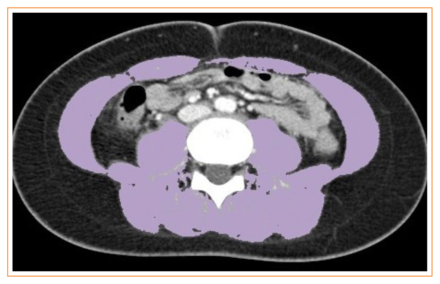
Sarcopenia is defined as the loss of skeletal muscle mass and is associated with negative clinical outcomes. This study aimed to establish sex-specific cutoff values for the skeletal muscle area (SMA) and skeletal muscle index (SMI) at the third lumbar vertebral (L3) level using computed tomography (CT) imaging to identify sarcopenia in healthy Korean liver donors.
Methods
This retrospective study included 659 healthy liver donors (408 men and 251 women) aged 20 to 60 years who had undergone abdominal CT examinations between January 2017 and December 2018. Assessment of body composition was performed with an automated segmentation technique using a deep-learning system. Sex-specific SMA and SMI distributions were assessed, and cutoff values for determining sarcopenia were defined as values at either two standard deviations (SDs) below the mean reference value or below the fifth percentile.
Results
Using the SD definition, cutoff values for SMA and SMI were 117.04 cm2 and 39.33 cm2/m2, respectively, in men and 71.39 cm2 and 27.77 cm2/m2, respectively, in women. Using the fifth percentile definition, cutoff values for SMA and SMI were 126.88 cm2 and 40.96 cm2/m2, respectively, in men and 78.85 cm2 and 30.60 cm2/m2, respectively, in women.
Conclusion
Our data provide sex-specific cutoff values for the SMA and SMI at the L3 level measured by CT imaging in a healthy Korean population, which may be applicable for identifying sarcopenia in this population. -
Citations
Citations to this article as recorded by
- Myosteatosis is associated with poor survival after kidney transplantation: a large retrospective cohort validation
Jie Chen, Yue Li, Chengjie Li, Turun Song
Abdominal Radiology.2024; 49(4): 1210. CrossRef - The effect of biological agent on body composition in patients with Crohn’s disease
Eun Jeong Choi, Dong Hoon Baek, Hong Sub Lee, Geun Am Song, Tae Oh Kim, Yong Eun Park, Chang Min Lee, Jong Hoon Lee
BMC Gastroenterology.2023;[Epub] CrossRef - The Association between the L3 Skeletal Muscle Index Derived from Computed Tomography and Clinical Outcomes in Patients with Urinary Tract Infection in the Emergency Department
Jinjoo An, Seung Pill Choi, Jae Hun Oh, Jong Ho Zhu, Sung Wook Kim, Soo Hyun Kim
Journal of Clinical Medicine.2023; 12(15): 5024. CrossRef - Validity of computed tomography defined body composition as a prognostic factor for functional outcome after kidney transplantation
Tim D. A. Swaab, Evelien E. Quint, Lisa B. Westenberg, Marcel Zorgdrager, Dorry L. Segev, Mara A. McAdams‐DeMarco, Stephan J. L. Bakker, Alain R. Viddeleer, Robert A. Pol
Journal of Cachexia, Sarcopenia and Muscle.2023; 14(6): 2532. CrossRef - Assessment of the Diaphragm Thickness Decrease in Critically Ill COVID-19 Patients: Could Computed Tomography Be of Aid Regarding Diaphragm Muscle Mass?
Oana-Elena Branea, Sanda Maria Copotoiu, Diana Andreea Becica, AnaMaria Romina Budeanu, Razvan Gabriel Budeanu, Mihai Emanuel Becica, Dragos Constantin Cucoranu, Septimiu Voidazan, Monica Chis, Alexandra Elena Lazar
Cureus.2023;[Epub] CrossRef - Clinical implication of thoracic skeletal muscle volume as a predictor of ventilation-weaning failure in brain-injured patients: A retrospective observational study
Jimi Oh, Hyun Lim, Chang Won Jeong, Min Su Kim, Jinseok Lee, Wu Seong Kang, Ui Ri An, Joo Un Park, Youngick Ahn, Youe Ree Kim, Chul Park
Medicine.2023; 102(43): e35847. CrossRef - Estimation of Muscle Mass Using Creatinine/Cystatin C Ratio in Japanese Community-Dwelling Older People
Hiroshi Kusunoki, Yasuharu Tabara, Shotaro Tsuji, Yosuke Wada, Kayoko Tamaki, Koutatsu Nagai, Masako Itoh, Kyoko Sano, Manabu Amano, Hatsuo Maeda, Hideyuki Sugita, Yoko Hasegawa, Hiromitsu Kishimoto, Soji Shimomura, Michiya Igase, Ken Shinmura
Journal of the American Medical Directors Association.2022; 23(5): 902.e21. CrossRef - Defining reference values for low skeletal muscle index at the L3 vertebra level based on computed tomography in healthy adults: A multicentre study
Ming Kong, Nan Geng, Ying Zhou, Ning Lin, Wenyan Song, Manman Xu, Shanshan Li, Yuetong Piao, Zuoqing Han, Rong Guo, Chao Yang, Nan Luo, Zhong Wang, Mengyuan Jiang, Lili Wang, Wanchun Qiu, Junfeng Li, Daimeng Shi, Rongkuan Li, Eddie C. Cheung, Yu Chen, Zho
Clinical Nutrition.2022; 41(2): 396. CrossRef - The Value of Artificial Intelligence-Assisted Imaging in Identifying Diagnostic Markers of Sarcopenia in Patients with Cancer
Ying-Tzu Huang, Yi-Shan Tsai, Peng-Chan Lin, Yu-Min Yeh, Ya-Ting Hsu, Pei-Ying Wu, Meng-Ru Shen, Zhongjie Shi
Disease Markers.2022; 2022: 1. CrossRef - Assessment of Muscle Quantity, Quality and Function
Bo Kyung Koo
Journal of Obesity & Metabolic Syndrome.2022; 31(1): 9. CrossRef - Computed Tomography-Derived Skeletal Muscle Radiodensity Is an Early, Sensitive Marker of Age-Related Musculoskeletal Changes in Healthy Adults
Yeon Woo Jung, Namki Hong, Joon Chae Na, Woong Kyu Han, Yumie Rhee
Endocrinology and Metabolism.2021; 36(6): 1201. CrossRef
- Myosteatosis is associated with poor survival after kidney transplantation: a large retrospective cohort validation

Review Articles
- Thyroid
- Percutaneous Adrenal Radiofrequency Ablation: A Short Review for Endocrinologists
- Byung Kwan Park
- Endocrinol Metab. 2020;35(4):750-755. Published online December 2, 2020
- DOI: https://doi.org/10.3803/EnM.2020.880
- 4,546 View
- 127 Download
- 9 Web of Science
- 8 Crossref
-
Abstract
PDFPubReader ePub
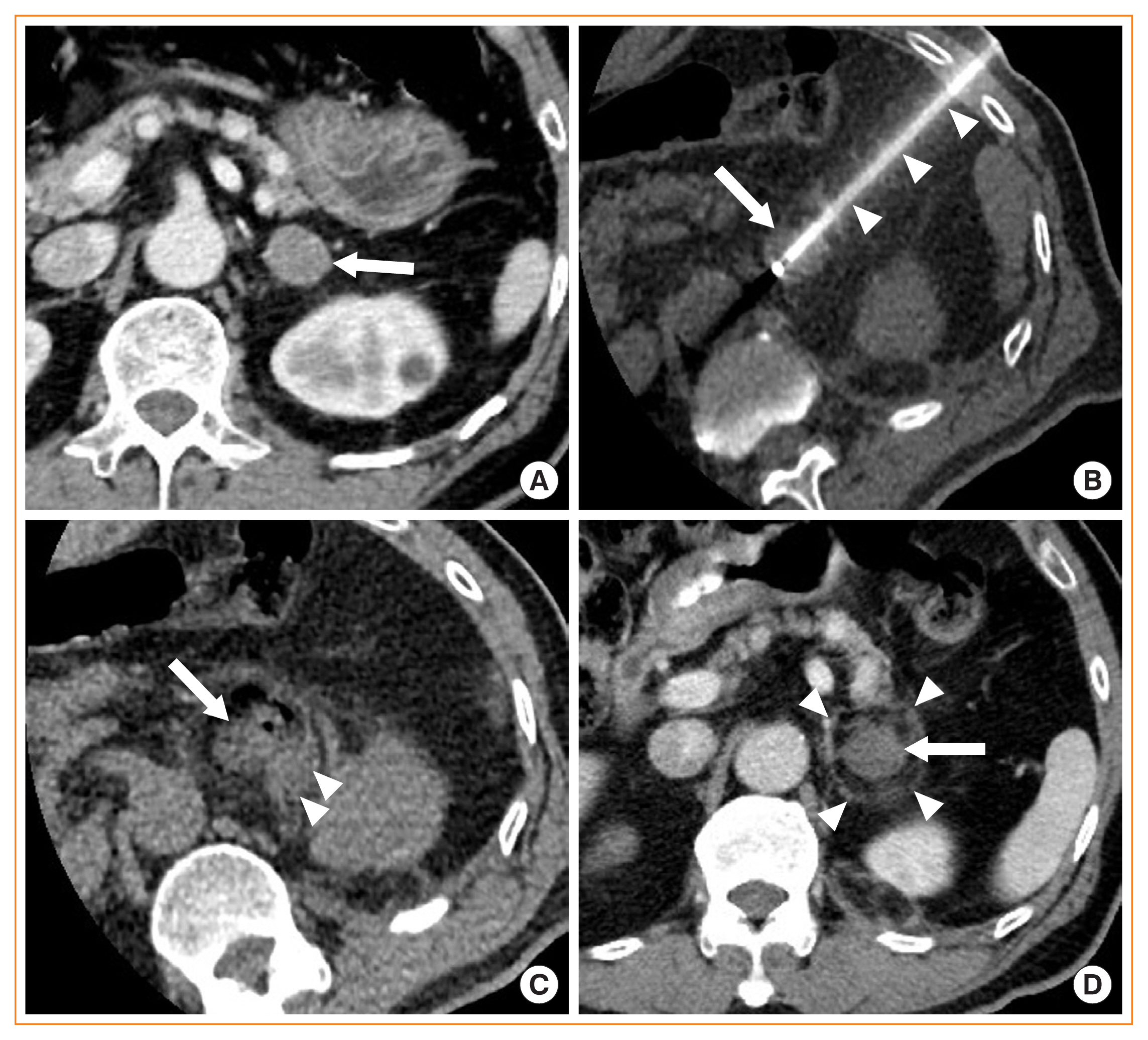
- Image-guided radiofrequency ablation (RFA) has been accepted as a minimally invasive treatment for adrenal tumors in patients who are unable to undergo adrenalectomy. Accordingly, this treatment has become more readily available for treating functioning or non-functioning adrenal masses. Thus, endocrinologists need a better understanding of percutaneous RFA of adrenal tumors. The purpose of this review is to briefly describe the basic mechanism of RFA, indications and contraindications, patient preparation prior to RFA, type of complications, how to avoid complications, RFA procedures, and treatment outcomes.
-
Citations
Citations to this article as recorded by- Clinical Features and Etiology of Recurrent Hypertension after Adrenalectomy
Xilan Dong, Qianhui Ling, Jin Bian, Yuehua Li, Mengjia Chen, Sufang Hao, Wenjun Ma, Huimin Zhang, Jun Cai, Ying Lou
Cardiovascular Innovations and Applications.2024;[Epub] CrossRef - Short-term outcome of adrenal radiofrequency ablation of adrenal cysts: a single-center experience
Shin Jeong Pak, Yu-mi Lee, Pyo Nyun Kim, Byung-Chang Kim, Jae Won Cho, Won Woong Kim, Tae-Yon Sung, Ki-wook Chung, Suck Joon Hong
Scientific Reports.2023;[Epub] CrossRef - Case report of trans-renal ablation procedures for a recurrent pheochromocytoma in von Hippel-Lindau disease
Byung Kwan Park
Precision and Future Medicine.2023; 7(2): 90. CrossRef - Efficacy and safety of radiofrequency ablation in the treatment of inoperable patients with pulmonary malignant nodules
Peng Qie, Xuejiao Xun, Xiaodong Nie, Qifan Yin, Hongshang Cui, Lijun Liu, Huien Wang
ANZ Journal of Surgery.2023; 93(12): 2969. CrossRef - 2023 Korean Endocrine Society Consensus Guidelines for the Diagnosis and Management of Primary Aldosteronism
Jeonghoon Ha, Jung Hwan Park, Kyoung Jin Kim, Jung Hee Kim, Kyong Yeun Jung, Jeongmin Lee, Jong Han Choi, Seung Hun Lee, Namki Hong, Jung Soo Lim, Byung Kwan Park, Jung-Han Kim, Kyeong Cheon Jung, Jooyoung Cho, Mi-kyung Kim, Choon Hee Chung
Endocrinology and Metabolism.2023; 38(6): 597. CrossRef - Minimally invasive techniques in benign and malignant adrenal tumors
Ahmet Bulent Dogrul, Omer Cennet, Anıl Hilmi Dincer
World Journal of Clinical Cases.2022; 10(35): 12812. CrossRef - Asian Conference on Tumor Ablation Guidelines for Adrenal Tumor Ablation
Byung Kwan Park, Masashi Fujimori, Shu-Huei Shen, Uei Pua
Endocrinology and Metabolism.2021; 36(3): 553. CrossRef - The role and place of mini-invasive methods of local tumor destruction in adrenal gland surgery
I.A. Kurganov, D.Yu. Bogdanov, S.I. Emelyanov, M.Sh. Mamistvalov
Endoskopicheskaya khirurgiya.2021; 27(6): 43. CrossRef
- Clinical Features and Etiology of Recurrent Hypertension after Adrenalectomy
- Adrenal gland
- Differential Diagnosis of Adrenal Mass Using Imaging Modality: Special Emphasis on F-18 Fluoro-2-Deoxy-D-Glucose Positron Emission Tomography/Computed Tomography
- Hong Je Lee, Jaetae Lee
- Endocrinol Metab. 2014;29(1):5-11. Published online March 14, 2014
- DOI: https://doi.org/10.3803/EnM.2014.29.1.5
- 4,105 View
- 30 Download
- 7 Web of Science
- 6 Crossref
-
Abstract
PDFPubReader
Adrenal incidentalomas are adrenal masses serendipitously detected during an imaging study performed for reasons unrelated to suspicion of adrenal disease. The incidence of adrenal incidentalomas has increased because of the widespread use of various imaging modalities. In oncology patients with adrenal incidentalomas, the characterization of the adrenal masses is challenging because nearly 50% of incidental adrenal masses are metastatic lesions that need special medical attention. Although unenhanced computed tomography (CT) densitometry, chemical shift magnetic resonance imaging (MRI), delayed contrast-enhanced CT and CT histogram analysis have been used as sensitive and specific modalities for differentiating benign from malignant adrenal masses, F-18 fluoro-2-deoxy-D-glucose positron emission tomography (F-18 FDG PET)/CT is a highly accurate imaging modality compared to CT or MRI, especially when these two imaging modalities are combined. In addition, a semiquantitative analysis using standardized uptake value ratio further improves the diagnostic accuracy of F-18 FDG PET/CT in differentiating benign from malignant adrenal masses. Thus, F-18 FDG PET/CT is very helpful for determining the best therapeutic management, especially for assessing the need for surgery.
-
Citations
Citations to this article as recorded by- The F-18 FDG PET/CT evaluation of the metastatic adrenal lesions of the non-lung cancer tumors compared with pathology results
Zehra Pınar Koç, Pınar Pelin Özcan, Emel Sezer, Kadir Eser, Tuba Kara
Egyptian Journal of Radiology and Nuclear Medicine.2022;[Epub] CrossRef - Dual time point [18F]Flurodeoxyglucose (FDG) Positron Emission Tomography (PET)/Computed Tomography (CT) with water gastric distension in differentiation between malignant and benign gastric lesions
Hussein Farghaly, Mohamed Alshareef, Abdullah Alqarni, Mohamed Sayed, Hatem Nasr
European Journal of Radiology Open.2020; 7: 100268. CrossRef - 18F-FDG PET/CT Imaging of Adrenal Liposarcoma
Rang Wang, Qiuping Fan, Rong Tian
Clinical Nuclear Medicine.2020; 45(7): 570. CrossRef - Characterization of adrenocortical tumors by 18F-FDG PET/CT: Does steroid hormone hypersecretion status modify the uptake pattern?
Nunzia Cinzia Paladino, Carole Guérin, Aoïfe Lowery, Andrea Attard, Wassim Essamet, Eveline Slotema, Isabelle Morange, Frédéric Castinetti, Thierry Brue, Anderson Loundou, David Taïeb, Frédéric Sebag
Surgical Oncology.2018; 27(2): 231. CrossRef - Articles in 'Endocrinology and Metabolism' in 2014
Won-Young Lee
Endocrinology and Metabolism.2015; 30(1): 47. CrossRef - Adrenal masses in oncology patients
Rodney H Reznek
Cancer Imaging.2014;[Epub] CrossRef
- The F-18 FDG PET/CT evaluation of the metastatic adrenal lesions of the non-lung cancer tumors compared with pathology results
 First
First Prev
Prev


