
Search
- Page Path
- HOME > Search
Original Article
- Miscellaneous
- DN200434 Inhibits Vascular Smooth Muscle Cell Proliferation and Prevents Neointima Formation in Mice after Carotid Artery Ligation
- Sudeep Kumar, Jonghwa Jin, Hyeon Young Park, Mi-Jin Kim, Jungwook Chin, Sungwoo Lee, Jina Kim, Jung-Guk Kim, Yeon-Kyung Choi, Keun-Gyu Park
- Endocrinol Metab. 2022;37(5):800-809. Published online September 28, 2022
- DOI: https://doi.org/10.3803/EnM.2022.1462

- 3,081 View
- 201 Download
- 1 Web of Science
- 1 Crossref
-
 Abstract
 PDF
PDF PubReader
  ePub
ePub - Background
Excessive proliferation and migration of vascular smooth muscle cells (VSMCs), which contributes to the development of occlusive vascular diseases, requires elevated mitochondrial oxidative phosphorylation to meet the increased requirements for energy and anabolic precursors. Therefore, therapeutic strategies based on blockade of mitochondrial oxidative phosphorylation are considered promising for treatment of occlusive vascular diseases. Here, we investigated whether DN200434, an orally available estrogen receptor-related gamma inverse agonist, inhibits proliferation and migration of VSMCs and neointima formation by suppressing mitochondrial oxidative phosphorylation.
Methods
VSMCs were isolated from the thoracic aortas of 4-week-old Sprague-Dawley rats. Oxidative phosphorylation and the cell cycle were analyzed in fetal bovine serum (FBS)- or platelet-derived growth factor (PDGF)-stimulated VSMCs using a Seahorse XF-24 analyzer and flow cytometry, respectively. A model of neointimal hyperplasia was generated by ligating the left common carotid artery in male C57BL/6J mice.
Results
DN200434 inhibited mitochondrial respiration and mammalian target of rapamycin complex 1 activity and consequently suppressed FBS- or PDGF-stimulated proliferation and migration of VSMCs and cell cycle progression. Furthermore, DN200434 reduced carotid artery ligation-induced neointima formation in mice.
Conclusion
Our data suggest that DN200434 is a therapeutic option to prevent the progression of atherosclerosis. -
Citations
Citations to this article as recorded by
- Jatrorrhizine inhibits Piezo1 activation and reduces vascular inflammation in endothelial cells
Tianying Hong, Xianmei Pan, Han Xu, Zhijuan Zheng, Lizhen Wen, Jing Li, Mingfeng Xia
Biomedicine & Pharmacotherapy.2023; 163: 114755. CrossRef
- Jatrorrhizine inhibits Piezo1 activation and reduces vascular inflammation in endothelial cells

Review Articles
- Bone Metabolism
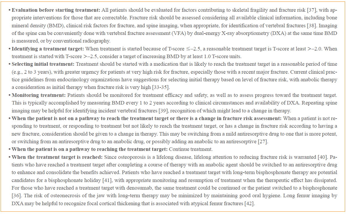
- Operationalizing Treat-to-Target for Osteoporosis
- E. Michael Lewiecki
- Endocrinol Metab. 2021;36(2):270-278. Published online March 24, 2021
- DOI: https://doi.org/10.3803/EnM.2021.970
- 5,622 View
- 299 Download
- 5 Web of Science
- 5 Crossref
-
Abstract
PDFPubReader ePub
- Treat-to-target (TTT) for osteoporosis is a concept for individualizing patient treatment decisions that focuses on achieving an acceptable level of fracture risk rather than response to treatment alone. While a response to treatment is essential in order to achieve an acceptable level of risk, it is not necessarily sufficient. Some patients have a good response to treatment yet remain at high level of fracture risk. Since there is no way to directly measure bone strength in patients treated for osteoporosis, a surrogate measurement must be used. Bone mineral density (BMD) is commonly used to select patients for treatment and has emerged as the most useful surrogate for assessing reduction of fracture risk after treatment is started. Recent large meta-regression studies have shown a robust correlation between larger increases in BMD with treatment and greater reductions in fracture risk. Application of TTT for osteoporosis involves assessing fracture risk before starting treatment and initiating treatment with an agent that is most likely to reduce fracture risk to an acceptable level, represented by a target BMD T-score, over a reasonable period of time. This review offers suggestions for implementing TTT for osteoporosis in clinical practice and managing patients who fail or succeed in reaching the target. More study is needed to fully validate the use of TTT for osteoporosis for initiating and modifying treatments to reduce fracture risk.
-
Citations
Citations to this article as recorded by- Treatment sequencing using the dual amylin and calcitonin receptor agonist KBP-336 and semaglutide results in durable weight loss
Anna Thorsø Larsen, Morten A. Karsdal, Kim Henriksen
European Journal of Pharmacology.2023; 954: 175837. CrossRef - Osteoporosis: Spotlight on current approaches to pharmacological treatment
Dilşad Sindel
Turkish Journal of Physical Medicine and Rehabilitation.2023; 69(2): 140. CrossRef - Postmenopausal Osteoporosis
Caren G. Solomon, Marcella Donovan Walker, Elizabeth Shane
New England Journal of Medicine.2023; 389(21): 1979. CrossRef - Prevalence and Risk Factors of T-Score Spine-Hip Discordance in Patients with Osteoporotic Vertebral Compression Fracture
Byung-Ho Yoon, Ho Won Kang, Su Min Kim, Young Do Koh
Journal of Bone Metabolism.2022; 29(1): 43. CrossRef - Pharmacological treatment of osteoporosis: 2022 update
Yunkyung Jeon, In-Joo Kim
Journal of the Korean Medical Association.2022; 65(4): 241. CrossRef
- Treatment sequencing using the dual amylin and calcitonin receptor agonist KBP-336 and semaglutide results in durable weight loss
- Acute Hyperglycemia Associated with Anti-Cancer Medication
- Yul Hwangbo, Eun Kyung Lee
- Endocrinol Metab. 2017;32(1):23-29. Published online March 20, 2017
- DOI: https://doi.org/10.3803/EnM.2017.32.1.23
- 6,324 View
- 132 Download
- 41 Web of Science
- 44 Crossref
-
Abstract
PDFPubReader
Hyperglycemia during chemotherapy occurs in approximately 10% to 30% of patients. Glucocorticoids and L-asparaginase are well known to cause acute hyperglycemia during chemotherapy. Long-term hyperglycemia is also frequently observed, especially in patients with hematologic malignancies treated with L-asparaginase-based regimens and total body irradiation. Glucocorticoid-induced hyperglycemia often develops because of increased insulin resistance, diminished insulin secretion, and exaggerated hepatic glucose output. Screening strategies for this condition include random glucose testing, hemoglobin A1c testing, oral glucose loading, and fasting plasma glucose screens. The management of hyperglycemia starts with insulin or sulfonylurea, depending on the type, dose, and delivery of the glucocorticoid formulation. Mammalian target of rapamycin (mTOR) inhibitors are associated with a high incidence of hyperglycemia, ranging from 13% to 50%. Immunotherapy, such as anti-programmed death 1 (PD-1) antibody treatment, induces hyperglycemia with a prevalence of 0.1%. The proposed mechanism of immunotherapy-induced hyperglycemia is an autoimmune process (insulitis). Withdrawal of the PD-1 inhibitor is the primary treatment for severe hyperglycemia. The efficacy of glucocorticoid therapy is not fully established and the decision to resume PD-1 inhibitor therapy depends on the severity of the hyperglycemia. Diabetic patients should achieve optimized glycemic control before initiating treatment, and glucose levels should be monitored periodically in patients initiating mTOR inhibitor or PD-1 inhibitor therapy. With regard to hyperglycemia caused by anti-cancer therapy, frequent monitoring and proper management are important for promoting the efficacy of anti-cancer therapy and improving patients' quality of life.
-
Citations
Citations to this article as recorded by- Bridging the Gap: Pancreas Tissue Slices From Organ and Tissue Donors for the Study of Diabetes Pathogenesis
Christian M. Cohrs, Chunguang Chen, Mark A. Atkinson, Denise M. Drotar, Stephan Speier
Diabetes.2024; 73(1): 11. CrossRef - Colorectal Cancer and Subsequent Diabetes Risk: A Population-Based Cohort Study in Taiwan
Hsin-Yin Hsu, Yih-Jong Chern, Min-Shu Hsu, Tzu-Lin Yeh, Ming-Chieh Tsai, Jing-Rong Jhuang, Cheng-Tzu Hsieh, Chun-Ju Chiang, Wen-Chung Lee, Lee-Ching Hwang, Kuo-Liong Chien
The Journal of Clinical Endocrinology & Metabolism.2024;[Epub] CrossRef - Assessment of metabolic syndrome parameters in pediatric acute lymphoblastic leukemia survivors
Ömer Kartal, Orhan Gürsel
Indian Journal of Cancer.2023; 60(3): 325. CrossRef - Increased risk of incident diabetes after therapy with immune checkpoint inhibitor compared with conventional chemotherapy: A longitudinal trajectory analysis using a tertiary care hospital database
Minyoung Lee, Kyeongseob Jeong, Yu Rang Park, Yumie Rhee
Metabolism.2023; 138: 155311. CrossRef - Analysis of the Incidence of Type 2 Diabetes, Requirement of Insulin Treatment, and Diabetes-Related Complications among Patients with Cancer
Su Jung Lee, Chulho Kim, Hyunjae Yu, Dong-Kyu Kim
Cancers.2023; 15(4): 1094. CrossRef - A Case of Pembrolizumab-Induced Diabetic Ketoacidosis and Hyperthyroidism in a Patient With Recurrent Esophageal Adenocarcinoma
Jonathan Salangsang, Surendra Sapkota, Sanjeev Kharel, Prakash Gupta, Abhishek Kalla
Cureus.2023;[Epub] CrossRef - Link between Blood Cell-Associated Inflammatory Indices and Chemotherapy-Induced Hyperglycemia in Women Affected with Breast Cancer: Clinical Studies
Krishna Prasad, Suresh Rao, Sanath Kumar Hegde, Thomas George, Rhea Katherine D'souza, Sucharitha Suresh, Manjeshwar Shrinath Baliga
South Asian Journal of Cancer.2023; 12(02): 118. CrossRef - Insulin resistance in patients with cancer: a systematic review and meta-analysis
Joan M. Màrmol, Michala Carlsson, Steffen H. Raun, Mia K. Grand, Jonas Sørensen, Louise Lang Lehrskov, Erik A. Richter, Ole Norgaard, Lykke Sylow
Acta Oncologica.2023; 62(4): 364. CrossRef - Metabolic obesity phenotypes and obesity‐related cancer risk in the National Health and Nutrition Examination Survey
Maci Winn, Prasoona Karra, Heinz Freisling, Marc J. Gunter, Benjamin Haaland, Michelle L. Litchman, Jennifer A. Doherty, Mary C. Playdon, Sheetal Hardikar
Endocrinology, Diabetes & Metabolism.2023;[Epub] CrossRef - Effects of Vitamin Intake on Blood Glucose in Cancer Patients Undergoing Chemotherapy: Quantitative and Descriptive Research
Ji Yeong Kim, Kyung Hee Lim
Korean Journal of Adult Nursing.2023; 35(2): 148. CrossRef - Oncologists’ responsibility, comfort, and knowledge managing hyperglycemia in patients with cancer undergoing chemotherapy: a cross sectional study
Teresa M. Salgado, Rotana M. Radwan, Erin Hickey Zacholski, Emily Mackler, Tonya M. Buffington, Kerri T. Musselman, William J. Irvin, Jennifer M. Perkins, Trang N. Le, Dave L. Dixon, Karen B. Farris, Vanessa B. Sheppard, Resa M. Jones
Supportive Care in Cancer.2023;[Epub] CrossRef - Diabetes management in cancer patients. An Italian Association of Medical Oncology, Italian Association of Medical Diabetologists, Italian Society of Diabetology, Italian Society of Endocrinology and Italian Society of Pharmacology multidisciplinary conse
N. Silvestris, T. Franchina, M. Gallo, A. Argentiero, A. Avogaro, G. Cirino, A. Colao, R. Danesi, G. Di Cianni, S. D’Oronzo, A. Faggiano, S. Fogli, D. Giuffrida, S. Gori, N. Marrano, R. Mazzilli, M. Monami, M. Montagnani, L. Morviducci, A. Natalicchio, A.
ESMO Open.2023; 8(6): 102062. CrossRef - Prognostic Factors for Hyperglycemia in Patients Receiving Chemotherapy
Jiyeong Kim, Kyung Hee Lim
Cancer Nursing.2023;[Epub] CrossRef - Severe Insulin Resistance in a Patient Treated With Nivolumab and Brentuximab-Vedotin for Hodgkin Lymphoma
Elif Tama, Meghan Black, Muhamad Alhaj Moustafa, Maria D Hurtado
JCEM Case Reports.2023;[Epub] CrossRef - A New Hypothesis Describing the Pathogenesis of Oral Mucosal Injury Associated with the Mammalian Target of Rapamycin (mTOR) Inhibitors
Stephen T. Sonis, Alessandro Villa
Cancers.2023; 16(1): 68. CrossRef - Management of Phosphatidylinositol-3-Kinase Inhibitor-Associated Hyperglycemia
Marcus D. Goncalves, Azeez Farooki
Integrative Cancer Therapies.2022; 21: 153473542110731. CrossRef - Metformin Induced Cognitive Impairment and Neuroinflammation in CMF-Treated Rats
Ahmad H. Alhowai, Yasser Almogbel, Ahmed A.H. Abdel, Maha A. Aldubay, Hani A. Alfheeaid, Shatha G. Felemban, Sridevi Chigurupat, Ibrahim F. Alharbi, Hindi S. Alharbi
International Journal of Pharmacology.2022; 18(2): 228. CrossRef - Glucose Influences the Response of Glioblastoma Cells to Temozolomide and Dexamethasone
Anna M Bielecka-Wajdman, Tomasz Ludyga, Daria Smyk, Wojciech Smyk, Magdalena Mularska, Patrycja Świderek, Wojciech Majewski, Christina Susanne Mullins, Michael Linnebacher, Ewa Obuchowicz
Cancer Control.2022; 29: 107327482210754. CrossRef - Cardiometabolic Comorbidities in Cancer Survivors
Leah L. Zullig, Anthony D. Sung, Michel G. Khouri, Shelley Jazowski, Nishant P. Shah, Andrea Sitlinger, Dan V. Blalock, Colette Whitney, Robin Kikuchi, Hayden B. Bosworth, Matthew J. Crowley, Karen M. Goldstein, Igor Klem, Kevin C. Oeffinger, Susan Dent
JACC: CardioOncology.2022; 4(2): 149. CrossRef - Glucose deprivation reduces proliferation and motility, and enhances the anti-proliferative effects of paclitaxel and doxorubicin in breast cell lines in vitro
Maitham A. Khajah, Sarah Khushaish, Yunus A. Luqmani, Yi-Hsien Hsieh
PLOS ONE.2022; 17(8): e0272449. CrossRef - Gemigliptin exerts protective effects against doxorubicin-induced hepatotoxicity by inhibiting apoptosis via the regulation of fibroblast growth factor 21 expression
Kyeong-Min Lee, Yeo Jin Hwang, Gwon-Soo Jung
Biochemical and Biophysical Research Communications.2022; 626: 135. CrossRef - Glucocorticoid-Induced Hyperglycemia in Oncologic Outpatients: A Narrative Review Using the Quadruple Aim Framework
Ihab Kandil, Erin Keely
Canadian Journal of Diabetes.2022; 46(7): 730. CrossRef - Real-World Experience of Monitoring Practice of Endocrinopathies Associated with the Use of Novel Targeted Therapies among Patients with Solid Tumors
Atika AlHarbi, Majed Alshamrani, Mansoor Khan, Abdelmajid Alnatsheh, Mohammed Aseeri
Medical Sciences.2022; 10(4): 65. CrossRef - The Assessment of the Hypothalamic-Pituitary-Adrenal Axis After Oncological Treatment in Pediatric Patients with Acute Lymphoblastic Leukemia
Barbara Hull, Anna Wedrychowicz, Magdalena Ossowska, Aleksandra Furtak, Joanna Badacz, Szymon Skoczeń, Jerzy B. Starzyk
Journal of Clinical Research in Pediatric Endocrinology.2022; 14(4): 393. CrossRef - Targeting Mitochondria and Oxidative Stress in Cancer- and Chemotherapy-Induced Muscle Wasting
Joshua R. Huot, Dryden Baumfalk, Aridai Resendiz, Andrea Bonetto, Ashley J. Smuder, Fabio Penna
Antioxidants & Redox Signaling.2022;[Epub] CrossRef - Onkodiabetológia III.
Róbert János Bánhegyi, Blanka Veréb, Andrea Gazdag, Beatrix Rácz, Róbert Wagner, Norbert Fülöp, Béla Pikó
Orvosi Hetilap.2022; 163(41): 1614. CrossRef - Economic burden of diabetes among medicare beneficiaries with cancer
Cassidi C McDaniel, F Ellen Loh, Devan M Rockwell, Courtney P McDonald, Chiahung Chou
Journal of Pharmaceutical Health Services Research.2021; 12(2): 142. CrossRef - The prognostic outcome of ‘type 2 diabetes mellitus and breast cancer’ association pivots on hypoxia-hyperglycemia axis
Ilhaam Ayaz Durrani, Attya Bhatti, Peter John
Cancer Cell International.2021;[Epub] CrossRef - Increased risk of diabetes in cancer survivors: a pooled analysis of 13 population-based cohort studies
Y. Xiao, H. Wang, Y. Tang, J. Yan, L. Cao, Z. Chen, Z. Shao, Z. Mei, Z. Jiang
ESMO Open.2021; 6(4): 100218. CrossRef - Sublethal concentrations of high glucose prolong mitotic arrest in a spindle assembly checkpoint activity dependent manner in budding yeast
Pinar B. Thomas, Elif E. Cavusoglu, Nur Kaluc
Biologia.2021; 76(12): 3883. CrossRef - Hyperglycemia in Childhood Acute Lymphoblastic Leukemia During Induction Chemotherapy
Nengcy Erlina Tasik Rerung, Andi Cahyadi, Nur Rochmah, Maria Christina Shanty Larasati, Mia Ratwita Andarsini, Muhammad Faizi, IDG Ugrasena, Bambang Permono
MEDICINUS.2021; 34(1): 18. CrossRef - Effect of Acute Chemotherapy on Glucose Levels in Rats
Ahmad H. Alhowail, Gena S. Alfawzan, Maha A. Aldubayan, Lolwah S. Alsalam
International Journal of Pharmacology.2020; 16(3): 276. CrossRef - Predictors of Disease Progression or Performance Status Decline in Patients Undergoing Neoadjuvant Therapy for Localized Pancreatic Head Adenocarcinoma
Alessandro Paniccia, Ana L. Gleisner, Mazen S. Zenati, Amr I. Al Abbas, Jae Pil Jung, Nathan Bahary, Kenneth K. W. Lee, David Bartlett, Melissa E. Hogg, Herbert J. Zeh, Amer H. Zureikat
Annals of Surgical Oncology.2020; 27(8): 2961. CrossRef - Fasting to enhance Cancer treatment in models: the next steps
Jing Zhang, Yanlin Deng, Bee Luan Khoo
Journal of Biomedical Science.2020;[Epub] CrossRef - Concurrent diabetic ketoacidosis and pancreatitis in Paediatric acute lymphoblastic leukemia receiving L-asparaginase
Patel Zeeshan Jameel, Sham Lohiya, Amol Dongre, Sachin Damke, Bhavana B. Lakhkar
BMC Pediatrics.2020;[Epub] CrossRef - Hyperglycemia during Adjuvant Chemotherapy as a Prognostic Factor in Breast Cancer Patients without Diabetes
Ha Rim Ahn, Sang Yull Kang, Hyun Jo Youn, Sung Hoo Jung
Journal of Breast Cancer.2020; 23(4): 398. CrossRef - Side effects of adjuvant chemotherapy and their impact on outcome in elderly breast cancer patients: a cohort study
Valentina Zanuso, Vittorio Fregoni, Lorenzo Gervaso
Future Science OA.2020; : FSO617. CrossRef - Incidence of Hyperglycemia/Secondary Diabetes in Women who have Undergone Curative Chemotherapy for Breast Cancer: First Study from India
Suresh Rao, Krishna Prasad, Soniya Abraham, Thomas George, Supreeth Kakkaje Chandran, Manjeshwar Shrinath Baliga
South Asian Journal of Cancer.2020; 09(03): 130. CrossRef - Hyperglycemic ADR Distribution of Doxorubicin From VigiBase
Jincheng Yang, Jun Yang
American Journal of Therapeutics.2019; 26(3): e428. CrossRef - Hyperglycemia During Childhood Cancer Therapy: Incidence, Implications, and Impact on Outcomes
Allison Grimes, Ashraf Mohamed, Jenna Sopfe, Rachel Hill, Jane Lynch
JNCI Monographs.2019; 2019(54): 132. CrossRef - Steroid-induced diabetes in cancer patients
Gemma Dinn
Journal of Prescribing Practice.2019; 1(12): 610. CrossRef - Risk Factors for Doxorubicin-Induced Serious Hyperglycaemia-Related Adverse Drug Reactions
Jincheng Yang, Yu Wang, Kang Liu, Wen Yang, Jianying Zhang
Diabetes Therapy.2019; 10(5): 1949. CrossRef - Effect of glucose and palmitate environment on proliferation and migration of PC3‐prostate cancer cells
Lívia Prometti Rezende, Maria Raquel Unterkircher Galheigo, Breno Costa Landim, Amanda Rodrigues Cruz, Françoise Vasconcelos Botelho, Renata Graciele Zanon, Rejane Maira Góes, Daniele Lisboa Ribeiro
Cell Biology International.2019; 43(4): 373. CrossRef - Incidence of Diabetes After Cancer Development
Yul Hwangbo, Danbee Kang, Minwoong Kang, Saemina Kim, Eun Kyung Lee, Young Ae Kim, Yoon Jung Chang, Kui Son Choi, So-Youn Jung, Sang Myung Woo, Jin Seok Ahn, Sung Hoon Sim, Yun Soo Hong, Roberto Pastor-Barriuso, Eliseo Guallar, Eun Sook Lee, Sun-Young Kon
JAMA Oncology.2018; 4(8): 1099. CrossRef
- Bridging the Gap: Pancreas Tissue Slices From Organ and Tissue Donors for the Study of Diabetes Pathogenesis
- Thyroid
- Recent Updates on the Management of Medullary Thyroid Carcinoma
- Bo Hyun Kim, In Joo Kim
- Endocrinol Metab. 2016;31(3):392-399. Published online August 26, 2016
- DOI: https://doi.org/10.3803/EnM.2016.31.3.392
- 4,725 View
- 59 Download
- 32 Web of Science
- 28 Crossref
-
Abstract
PDFPubReader
Medullary thyroid carcinoma (MTC) is a rare neuroendocrine tumor derived from the thyroid C cells producing calcitonin. MTC accounts for 0.6% of all thyroid cancers and incidence of MTC increased steadily between 1997 and 2011 in Korea. It occurs either sporadically or in a hereditary form based on germline rearranged during transfection (
RET ) mutations. MTC can be cured only by complete resection of the thyroid tumor and any loco-regional metastases. The most appropriate treatment is still less clear in patients with residual or recurrent disease after initial surgery or those with distant metastases because most patients even with metastatic disease have indolent courses with slow progression for several years and MTC is not responsive to either radioactive iodine therapy or thyroid-stimulating hormone suppression. Recently, two tyrosine kinase inhibitors (TKIs), vandetanib and cabozantinib, are approved for use in patients with advanced, metastatic or progressive MTC. In this review, we summarize the current approach according to revised American Thyroid Association guidelines and recent advances in systemic treatment such as TKIs for patients with persistent or recurrent MTC after surgery.-
Citations
Citations to this article as recorded by- [68Ga]Ga-DOTA-FAPI-04 PET/CT depicts metastases from medullary thyroid cancer that [68Ga]Ga-DOTATOC PET/CT missed
Akram Al-Ibraheem, Salem Fandi Alyasjeen, Ahmed Saad Abdlkadir, Areej Abu Sheikha
European Journal of Nuclear Medicine and Molecular Imaging.2023; 50(13): 4112. CrossRef - Molecular Mechanism and Clinical Application of Thyroid Cancer
·如孜 卡地力艳
Advances in Clinical Medicine.2023; 13(12): 20543. CrossRef - Research Progress of the Molecular Mechanism of Antithyroid Cancer Activity
of Shikonin
Chunguang Sun, Lin Liao
Current Molecular Pharmacology.2023;[Epub] CrossRef - Multiple endocrine neoplasia 2: an overview
B Saravana-Bawan, JD Pasternak
Therapeutic Advances in Chronic Disease.2022; 13: 204062232210792. CrossRef - Total thyroidectomy vs thyroid lobectomy for localized medullary thyroid cancer in adults: A propensity-matched survival analysis
Weili Liang, Jinyuan Shi, Hui Zhang, Guixu Lv, Tiantian Wang, Yong Wang, Bin Lv, Luchuan Li, Qingdong Zeng, Lei Sheng
Surgery.2022; 172(5): 1385. CrossRef - Naphthoquinones and derivatives as potential anticancer agents: An updated review
Md Mominur Rahman, Md Rezaul Islam, Shopnil Akash, Sheikh Shohag, Limon Ahmed, Fatema Akter Supti, Abdur Rauf, Abdullah S.M. Aljohani, Waleed Al Abdulmonem, Anees Ahmed Khalil, Rohit Sharma, Muthu Thiruvengadam
Chemico-Biological Interactions.2022; 368: 110198. CrossRef - Metformin Decreases Serum Thyroglobulin Concentration in Nonmedullary Thyroid Carcinoma
Celina Caetano, Paola Tabaro Pico, Charan Singh, Beatriz Tendler, Diana M Malchoff, Carl D Malchoff
Journal of the Endocrine Society.2022;[Epub] CrossRef - Long non-coding RNA LINC00488 facilitates thyroid cancer cell progression through miR-376a-3p/PON2
Fuyuan Xie, Longgen Li, Yuting Luo, Rensheng Chen, Jinhong Mei
Bioscience Reports.2021;[Epub] CrossRef - Decision Making When Cancer Becomes Chronic: Needs Assessment for a Web-Based Medullary Thyroid Carcinoma Patient Decision Aid
Danielle Shojaie, Aubri S Hoffman, Ruth Amaku, Maria E Cabanillas, Julie Ann Sosa, Steven G Waguespack, Mark E Zafereo, Mimi I Hu, Elizabeth E Grubbs
JMIR Formative Research.2021; 5(7): e27484. CrossRef - Long Non-coding RNA RUNDC3A-AS1 Promotes Lung Metastasis of Thyroid Cancer via Targeting the miR-182-5p/ADAM9
Dawei Ma, Yan Zhu, Xiao Zhang, Jia Zhang, Wei Chen, Xinyuan Chen, Yichun Qian, Yanbin Zhao, Tingting Hu, Zhangyu Yao, Wei Zhao, Yuan Zhang, Fangzhou Liu
Frontiers in Cell and Developmental Biology.2021;[Epub] CrossRef - Effects of marital status on survival of medullary thyroid cancer stratified by age
Lei Ai, Ning Li, Hai‐Long Tan, Bo Wei, Ya‐Xin Zhao, Pei Chen, Hui‐Yu Hu, Mian Liu, Deng‐Jie Ou‐Yang, Zi‐en Qin, Peng Huang, Shi Chang
Cancer Medicine.2021; 10(24): 8829. CrossRef - The prognostic value of lymph node ratio in Medullary thyroid carcinoma: A multi-center study
Tal Rozenblat, Dania Hirsch, Eyal Robenshtok, Simona Grozinsky-Glasberg, David J. Gross, Haggi Mazeh, Carlos Benbassat, Orit Twito, Sigal Levy, Aviram Mizrachi, Thomas Shpitzer, Gideon Bachar
European Journal of Surgical Oncology.2020; 46(11): 2023. CrossRef Multiple Auer Rods in Fine-Needle Aspiration Smears of Medullary Thyroid Carcinoma: An Unusual Finding
Maryam Mohammadnia Avval, Perikala Vijayananda Kumar, Fereydoon Dehghani
International Medical Case Reports Journal.2020; Volume 13: 85. CrossRef- Lymph Node Metastases of Medullary Thyroid Cancer: Role of Calcitonin in the Washout Fluid of Fine-Needle Aspiration
Bernardo Marques, Nuno Cunha, Raquel G. Martins, Ana Rita Elvas, Joana Couto, Jacinta Santos, Teresa Martins, Ana Paula Moniz, Olga Ilhéu, Frederico Valido, Fernando Rodrigues
International Journal of Endocrinology.2020; 2020: 1. CrossRef - Efficacy and Safety of Vandetanib in Progressive and Symptomatic Medullary Thyroid Cancer: Post Hoc Analysis From the ZETA Trial
Michael C. Kreissl, Lars Bastholt, Rossella Elisei, Robert Haddad, Ole Hauch, Barbara Jarząb, Bruce Robinson, Raffaella Colzani, Meredith Foster, Richard Weiss, Martin Schlumberger
Journal of Clinical Oncology.2020; 38(24): 2773. CrossRef - Medullary thyroid cancer: epidemiological pattern and factors contributing to recurrence and metastasis
O Hamdy, S Awny, IH Metwally
The Annals of The Royal College of Surgeons of England.2020; 102(7): 499. CrossRef - Carcinoma medular de tiroides estadio iv y gestación
B. Sánchez Lechuga, C. López Tinoco, I. Gavilán Villarejo, F.J. Vílchez López, M. Aguilar Diosdado
Clínica e Investigación en Ginecología y Obstetricia.2019; 46(1): 35. CrossRef - Bildgebung beim medullären Schilddrüsenkarzinom
M. Uhrig, S. Delorme
Der Radiologe.2019; 59(11): 992. CrossRef - Recent Development of Nuclear Molecular Imaging in Thyroid Cancer
Huiting Liu, Xiaoqin Wang, Ran Yang, Wenbing Zeng, Dong Peng, Jason Li, Hu Wang
BioMed Research International.2018; 2018: 1. CrossRef - Radioguided hepatic resection with l -DOPA in metastatic medullary thyroid carcinoma
Javier López-Gómez, Sevastian Medina-Ornelas, Ma. Alejandra Salazar-Álvarez, Mario Álvarez-Bojórquez, Noel Zaragoza-Cruz, Javier Melchor-Ruan, Alethia Álvarez-Cano
Revista Española de Medicina Nuclear e Imagen Molecular (English Edition).2018; 37(4): 244. CrossRef - EF24 (a Curcumin Analog) and ZSTK474 Emphasize the Effect of Cabozantinib in Medullary Thyroid Cancer
Loris Bertazza, Francesca Sensi, Elisabetta Cavedon, Sara Watutantrige-Fernando, Simona Censi, Jacopo Manso, Federica Vianello, Eric Casal Ide, Maurizio Iacobone, Raffaele Pezzani, Caterina Mian, Susi Barollo
Endocrinology.2018; 159(6): 2348. CrossRef - Resección hepática radioguiada con 18 F-DOPA en un paciente con carcinoma medular de tiroides metastásico
J. López-Gómez, S. Medina-Ornelas, M.A. Salazar-Álvarez, M. Álvarez-Bojórquez, N. Zaragoza-Cruz, J. Melchor-Ruan, A. Álvarez-Cano
Revista Española de Medicina Nuclear e Imagen Molecular.2018; 37(4): 244. CrossRef - Genotypic characteristics and their association with phenotypic characteristics of hereditary medullary thyroid carcinoma in Korea
Kyong Yeun Jung, Seok-Mo Kim, Min Joo Kim, Sun Wook Cho, Bup-Woo Kim, Yong Sang Lee, Jong Ju Jeong, Kee-Hyun Nam, Woong Youn Chung, Kyu Eun Lee, Eun-Jae Chung, Hyo Jeong Kim, Do Joon Park, Myung-Whun Sung, Cheong Soo Park, Bo Youn Cho, Young Joo Park, Han
Surgery.2018; 164(2): 312. CrossRef - Thyroid cancer management
Francesco Perri, Antonio Giordano, Salvatore Pisconti, Franco Ionna, Maria G. Chiofalo, Francesco Longo, Davide Leopardo, Giuseppina Della Vittoria Scarpati, Luciano Pezzullo
Anti-Cancer Drugs.2018; 29(6): 483. CrossRef - Clinicopathological Significance and Prognosis of Medullary Thyroid Microcarcinoma: A Meta‐analysis
Jin Hwa Kim, Jung‐Soo Pyo, Won Jin Cho
World Journal of Surgery.2017; 41(10): 2551. CrossRef - A label-free electrochemical immunosensor based on gold nanoparticles and graphene oxide for the detection of tumor marker calcitonin
Nawal A. Alarfaj, Maha F. El-Tohamy
New Journal of Chemistry.2017; 41(19): 11029. CrossRef - Evaluation of in vitro and in vivo activity of a multityrosine kinase inhibitor, AL3810, against human thyroid cancer
Qin Xie, Hui Chen, Jing Ai, Ying-lei Gao, Mei-yu Geng, Jian Ding, Yi Chen
Acta Pharmacologica Sinica.2017; 38(11): 1533. CrossRef - Articles inEndocrinology and Metabolismin 2016
Won-Young Lee
Endocrinology and Metabolism.2017; 32(1): 62. CrossRef
- [68Ga]Ga-DOTA-FAPI-04 PET/CT depicts metastases from medullary thyroid cancer that [68Ga]Ga-DOTATOC PET/CT missed
- Obesity and Metabolism
- Understanding Metabolomics in Biomedical Research
- Su Jung Kim, Su Hee Kim, Ji Hyun Kim, Shin Hwang, Hyun Ju Yoo
- Endocrinol Metab. 2016;31(1):7-16. Published online March 16, 2016
- DOI: https://doi.org/10.3803/EnM.2016.31.1.7
- 7,036 View
- 127 Download
- 46 Web of Science
- 42 Crossref
-
Abstract
PDFPubReader
The term "omics" refers to any type of specific study that provides collective information on a biological system. Representative omics includes genomics, proteomics, and metabolomics, and new omics is constantly being added, such as lipidomics or glycomics. Each omics technique is crucial to the understanding of various biological systems and complements the information provided by the other approaches. The main strengths of metabolomics are that metabolites are closely related to the phenotypes of living organisms and provide information on biochemical activities by reflecting the substrates and products of cellular metabolism. The transcriptome does not always correlate with the proteome, and the translated proteome might not be functionally active. Therefore, their changes do not always result in phenotypic alterations. Unlike the genome or proteome, the metabolome is often called the molecular phenotype of living organisms and is easily translated into biological conditions and disease states. Here, we review the general strategies of mass spectrometry-based metabolomics. Targeted metabolome or lipidome analysis is discussed, as well as nontargeted approaches, with a brief explanation of the advantages and disadvantages of each platform. Biomedical applications that use mass spectrometry-based metabolomics are briefly introduced.
-
Citations
Citations to this article as recorded by- Urine metabolomic biomarkers for prediction of isolated fetal congenital heart defect
Perry Friedman, Ali Yilmaz, Zafer Ugur, Faryal Jafar, Amy Whitten, Ilyas Ustun, Onur Turkoglu, Stewart Graham, Ray Bahado Singh
The Journal of Maternal-Fetal & Neonatal Medicine.2024; 35(25): 6380. CrossRef - Metabolomics and Risk of Dementia: A Systematic Review of Prospective Studies
Ashley C Flores, Xinyuan Zhang, Penny M Kris-Etherton, Martin J Sliwinski, Greg C Shearer, Xiang Gao, Muzi Na
The Journal of Nutrition.2024; 154(3): 826. CrossRef - Associations of PFAS-related plasma metabolites with cholesterol and triglyceride concentrations
T. Schillemans, I.A. Bergdahl, K. Hanhineva, L. Shi, C. Donat-Vargas, J. Koponen, H. Kiviranta, R. Landberg, A. Åkesson, C. Brunius
Environmental Research.2023; 216: 114570. CrossRef - UPLC-Q-Exactive-based rats serum metabolomics for characterization of traditional Chinese medicine Natures and Flavors
Hong Wang, Ruofang Gao, Jing Liu, Shuang Zhang, Yunli Zhao, Zhiguo Yu
Journal of Ethnopharmacology.2023; 302: 115931. CrossRef - Artificial Neural Networks Coupled with MALDI-TOF MS Serum Fingerprinting To Classify and Diagnose Pathological Pain Subtypes in Preclinical Models
Meritxell Deulofeu, Eladia M. Peña-Méndez, Petr Vaňhara, Josef Havel, Lukáš Moráň, Lukáš Pečinka, Anna Bagó-Mas, Enrique Verdú, Victoria Salvadó, Pere Boadas-Vaello
ACS Chemical Neuroscience.2023; 14(2): 300. CrossRef - Metabolic signature of HepaRG cells exposed to ethanol and tumor necrosis factor alpha to study alcoholic steatohepatitis by LC–MS-based untargeted metabolomics
Elias Iturrospe, Rani Robeyns, Katyeny Manuela da Silva, Maria van de Lavoir, Joost Boeckmans, Tamara Vanhaecke, Alexander L. N. van Nuijs, Adrian Covaci
Archives of Toxicology.2023; 97(5): 1335. CrossRef - Integrating metabolomics and network pharmacology to assess the effects of quercetin on lung inflammatory injury induced by human respiratory syncytial virus
Ya-Lei Sun, Pei-Pei Zhao, Cheng-Bi Zhu, Ming-Chen Jiang, Xin-Min Li, Jia-Lei Tao, Chan-Chan Hu, Bin Yuan
Scientific Reports.2023;[Epub] CrossRef - Comparison of Plasma Metabolites From Patients With Non-Small Cell Lung Cancer by Erlotinib Treatment and Skin Rash
Won Kil Lee, Jisoo Myong, Eunbin Kwag, Younmin Shin, Ji Woong Son, Byong Chul Yoo, Byoung-Soo Kim, Hwa-Seung Yoo, Jeong June Choi
Integrative Cancer Therapies.2023;[Epub] CrossRef - Metabolomic Signatures of Exposure to Nitrate and Trihalomethanes in Drinking Water and Colorectal Cancer Risk in a Spanish Multicentric Study (MCC-Spain)
Jose A. Alcolea, Carolina Donat-Vargas, Anastasia Chrysovalantou Chatziioannou, Pekka Keski-Rahkonen, Nivonirina Robinot, Antonio José Molina, Pilar Amiano, Inés Gómez-Acebo, Gemma Castaño-Vinyals, Lea Maitre, Marc Chadeau-Hyam, Sonia Dagnino, Sibo Lucas
Environmental Science & Technology.2023; 57(48): 19316. CrossRef - Metabolic Signature of Ethanol-Induced Hepatotoxicity in HepaRG Cells by Liquid Chromatography–Mass Spectrometry-Based Untargeted Metabolomics
Elias Iturrospe, Katyeny Manuela da Silva, Rani Robeyns, Maria van de Lavoir, Joost Boeckmans, Tamara Vanhaecke, Alexander L.N. van Nuijs, Adrian Covaci
Journal of Proteome Research.2022; 21(4): 1153. CrossRef - Current State and Challenges of the Global Outcomes of Dental Caries Research in the Meta-Omics Era
Dina G. Moussa, Paras Ahmad, Tamer A. Mansour, Walter L. Siqueira
Frontiers in Cellular and Infection Microbiology.2022;[Epub] CrossRef - Current Knowledge in Skin Metabolomics: Updates from Literature Review
Alessia Paganelli, Valeria Righi, Elisabetta Tarentini, Cristina Magnoni
International Journal of Molecular Sciences.2022; 23(15): 8776. CrossRef - Metabolome-wide association study of the relationship between chlorpyrifos exposure and first trimester serum metabolite levels in pregnant Thai farmworkers
Donghai Liang, Jonathan Batross, Nancy Fiedler, Tippawan Prapamontol, Panrapee Suttiwan, Parinya Panuwet, Warangkana Naksen, Brittney O. Baumert, Volha Yakimavets, Youran Tan, Priya D'Souza, Ampica Mangklabruks, Supattra Sittiwang, Kristsanachai Kaewthit,
Environmental Research.2022; 215: 114319. CrossRef - Differential Plasma Metabolites between High- and Low-Grade Meningioma Cases
Gabriel A. Kurokawa, Pedro T. Hamamoto Filho, Jeany Delafiori, Aline F. Galvani, Arthur N. de Oliveira, Flávia L. Dias-Audibert, Rodrigo R. Catharino, Maria Inês M. C. Pardini, Marco A. Zanini, Estela de O. Lima, Adriana C. Ferrasi
International Journal of Molecular Sciences.2022; 24(1): 394. CrossRef - Plasma metabolites associated with exposure to perfluoroalkyl substances and risk of type 2 diabetes – A nested case-control study
Tessa Schillemans, Lin Shi, Carolina Donat-Vargas, Kati Hanhineva, Andreas Tornevi, Ingegerd Johansson, Jani Koponen, Hannu Kiviranta, Olov Rolandsson, Ingvar A. Bergdahl, Rikard Landberg, Agneta Åkesson, Carl Brunius
Environment International.2021; 146: 106180. CrossRef - Methionine transsulfuration pathway is upregulated in long-lived humans
Natàlia Mota-Martorell, Mariona Jové, Consuelo Borrás, Rebeca Berdún, Èlia Obis, Joaquim Sol, Rosanna Cabré, Irene Pradas, José Daniel Galo-Licona, Josep Puig, José Viña, Reinald Pamplona
Free Radical Biology and Medicine.2021; 162: 38. CrossRef - Investigating metabolism by mass spectrometry: From steady state to dynamic view
Matteo Audano, Silvia Pedretti, Simona Ligorio, Flavio Giavarini, Donatella Caruso, Nico Mitro
Journal of Mass Spectrometry.2021;[Epub] CrossRef - The Efficacy of Pilates on Urinary Incontinence in Korean Women: A Metabolomics Approach
Gyumin Kang, Haelim Lee, Malsoon Shin, Jaekwan Kim, Sungki Lee, Youngja Park
Metabolites.2021; 11(2): 118. CrossRef - Broad Metabolome Alterations Associated with the Intake of Oral Contraceptives Are Mediated by Cortisol in Premenopausal Women
Clara Eick, Johanna Klinger-König, Stephanie Zylla, Anke Hannemann, Kathrin Budde, Ann Kristin Henning, Maik Pietzner, Matthias Nauck, Henry Völzke, Hans J. Grabe, Johannes Hertel
Metabolites.2021; 11(4): 193. CrossRef - Nutritional Metabolomics and the Classification of Dietary Biomarker Candidates: A Critical Review
Talha Rafiq, Sandi M Azab, Koon K Teo, Lehana Thabane, Sonia S Anand, Katherine M Morrison, Russell J de Souza, Philip Britz-McKibbin
Advances in Nutrition.2021; 12(6): 2333. CrossRef - MSCAT: A Machine Learning Assisted Catalog of Metabolomics Software Tools
Jonathan Dekermanjian, Wladimir Labeikovsky, Debashis Ghosh, Katerina Kechris
Metabolites.2021; 11(10): 678. CrossRef - Defining NASH from a Multi-Omics Systems Biology Perspective
Lili Niu, Karolina Sulek, Catherine G. Vasilopoulou, Alberto Santos, Nicolai J. Wewer Albrechtsen, Simon Rasmussen, Florian Meier, Matthias Mann
Journal of Clinical Medicine.2021; 10(20): 4673. CrossRef - Defining Blood Plasma and Serum Metabolome by GC-MS
Olga Kiseleva, Ilya Kurbatov, Ekaterina Ilgisonis, Ekaterina Poverennaya
Metabolites.2021; 12(1): 15. CrossRef - Precision Medicine in Childhood Asthma: Omic Studies of Treatment Response
Javier Perez-Garcia, Esther Herrera-Luis, Fabian Lorenzo-Diaz, Mario González, Olaia Sardón, Jesús Villar, Maria Pino-Yanes
International Journal of Molecular Sciences.2020; 21(8): 2908. CrossRef - Metabolomic characterization of semen from asthenozoospermic patients using ultra‐high‐performance liquid chromatography–tandem quadrupole time‐of‐flight mass spectrometry
Li Li, Xinrui Hao, Hua Chen, Liyuan Wang, Aolei Chen, Xiaotian Song, Zhiyan Hu, Ying Su, Han Lin, Pei Fan
Biomedical Chromatography.2020;[Epub] CrossRef - Comparison of Metabolomic Profiles of Organs in Mice of Different Strains Based on SPME-LC-HRMS
Katarzyna Burlikowska, Iga Stryjak, Joanna Bogusiewicz, Bogumiła Kupcewicz, Karol Jaroch, Barbara Bojko
Metabolites.2020; 10(6): 255. CrossRef - Metabolomics and Machine Learning Approaches Combined in Pursuit for More Accurate Paracoccidioidomycosis Diagnoses
Estela de Oliveira Lima, Luiz Claudio Navarro, Karen Noda Morishita, Camila Mika Kamikawa, Rafael Gustavo Martins Rodrigues, Mohamed Ziad Dabaja, Diogo Noin de Oliveira, Jeany Delafiori, Flávia Luísa Dias-Audibert, Marta da Silva Ribeiro, Adriana Pardini
mSystems.2020;[Epub] CrossRef - Metabolomics in the study of spontaneous animal diseases
Helena Tran, Malcolm McConville, Panayiotis Loukopoulos
Journal of Veterinary Diagnostic Investigation.2020; 32(5): 635. CrossRef - Effect of Pioglitazone on Perihematomal Edema in Intracerebral Hemorrhage Mouse Model by Regulating NLRP3 Expression and Energy Metabolism
Hoon Kim, Jung Eun Lee, Hyun Ju Yoo, Jae Hoon Sung, Seung Ho Yang
Journal of Korean Neurosurgical Society.2020; 63(6): 689. CrossRef - Inborn Errors of Metabolism in the Era of Untargeted Metabolomics and Lipidomics
Ismail, Showalter, Fiehn
Metabolites.2019; 9(10): 242. CrossRef - Metabolomic profiles of induced pluripotent stem cells derived from patients with rheumatoid arthritis and osteoarthritis
Juryun Kim, Sunyoung Christina Kang, Na Eun Yoon, Yena Kim, Jinhyeok Choi, Narae Park, Hyerin Jung, Byung Hwa Jung, Ji Hyeon Ju
Stem Cell Research & Therapy.2019;[Epub] CrossRef - Metabolomics profiles of patients with Wilson disease reveal a distinct metabolic signature
Gaurav V. Sarode, Kyoungmi Kim, Dorothy A. Kieffer, Noreene M. Shibata, Tomas Litwin, Anna Czlonkowska, Valentina Medici
Metabolomics.2019;[Epub] CrossRef - Metabolomics study of the therapeutic mechanism of a Chinese herbal formula on collagen-induced arthritis mice
Zhen Jin, Ji-da Zhang, Xin Wu, Gang Cao
RSC Advances.2019; 9(7): 3716. CrossRef - Plasma sphingomyelins increase in pre-diabetic Korean men with abdominal obesity
Seung-Soon Im, Hyeon Young Park, Jong Cheol Shon, In-Sung Chung, Ho Chan Cho, Kwang-Hyeon Liu, Dae-Kyu Song, Kyoung Heon Kim
PLOS ONE.2019; 14(3): e0213285. CrossRef - Tissue‐Specific Metabolic Profiles After Prolonged Cardiac Arrest Reveal Brain Metabolome Dysfunction Predominantly After Resuscitation
Jaewoo Choi, Muhammad Shoaib, Tai Yin, Gautam Nayyar, Koichiro Shinozaki, Jan F. Stevens, Lance B. Becker, Junhwan Kim
Journal of the American Heart Association.2019;[Epub] CrossRef - Global serum metabolomics profiling of colorectal cancer (Review)
Nurul Amir Hashim, Sharaniza Ab‑Rahim, Leny Suddin, Mohd Ahmad Saman, Musalmah Mazlan
Molecular and Clinical Oncology.2019;[Epub] CrossRef - Metabolomic Profiling of Human Spermatozoa in Idiopathic Asthenozoospermia Patients Using Gas Chromatography-Mass Spectrometry
Kai Zhao, Jianzhong Zhang, Zhen Xu, Yue Xu, Aiming Xu, Wei Chen, Chenkui Miao, Shouyong Liu, Zengjun Wang, Ruipeng Jia
BioMed Research International.2018; 2018: 1. CrossRef - Articles inEndocrinology and Metabolismin 2016
Won-Young Lee
Endocrinology and Metabolism.2017; 32(1): 62. CrossRef - Investigation of the effects of storage and freezing on mixes of heavy‐labeled metabolite and amino acid standards
Rachel Culp‐Hill, Julie A. Reisz, Kirk C. Hansen, Angelo D'Alessandro
Rapid Communications in Mass Spectrometry.2017; 31(23): 2030. CrossRef - Beyond the paradigm: Combining mass spectrometry and nuclear magnetic resonance for metabolomics
Darrell D. Marshall, Robert Powers
Progress in Nuclear Magnetic Resonance Spectroscopy.2017; 100: 1. CrossRef - Untargeted metabolomics analysis of adipogenic transformation in OP9‐DL1 cells using liquid chromatography‐mass spectrometry: Implications for thymic adipogenesis
Jianxin Tan, Yajun Wang, Siliang Wang, Nannan Zhang, Simeng Wu, Zhe Yuan, Xike Zhu
Cell Biology International.2017; 41(4): 447. CrossRef - Gut Microbiota Profiling: Metabolomics Based Approach to Unravel Compounds Affecting Human Health
Pamela Vernocchi, Federica Del Chierico, Lorenza Putignani
Frontiers in Microbiology.2016;[Epub] CrossRef
- Urine metabolomic biomarkers for prediction of isolated fetal congenital heart defect
- Thyroid
- Radioiodine Therapy in Differentiated Thyroid Cancer: The First Targeted Therapy in Oncology
- June-Key Chung, Gi Jeong Cheon
- Endocrinol Metab. 2014;29(3):233-239. Published online September 25, 2014
- DOI: https://doi.org/10.3803/EnM.2014.29.3.233
- 4,934 View
- 75 Download
- 34 Web of Science
- 34 Crossref
-
Abstract
PDFPubReader
Iodide uptake across the membranes of thyroid follicular cells and cancer cells occurs through an active transport process mediated by the sodium-iodide symporter (NIS). The rat and human NIS-coding genes were cloned and identified in 1996. Evaluation of NIS gene and protein expression is critical for the management of thyroid cancer, and several approaches to increase NIS levels have been tried. Identification of the NIS gene has provided a means of expanding its role in radionuclide therapy and molecular target-specific theragnosis (therapy and diagnosis using the same molecular target). In this article, we describe the relationship between NIS expression and the thyroid carcinoma treatment using I-131 and alternative therapeutic approaches.
-
Citations
Citations to this article as recorded by- A Pilot Nonrandomized Controlled Trial Examining the Use of Artificial Tears on the Radioactivity of Tears After Radioactive Iodine Treatment for Thyroid Cancer
Irene T. Lee, Jared V. Grice, Xiangyu Ji, Qingxia Chen, Lindsay A. Bischoff, Aaron C. Jessop, Behin Barahimi, Francois Codere, Louise A. Mawn, Rachel K. Sobel
Thyroid®.2024; 34(1): 82. CrossRef - Research progress of bone metastases: From disease recognition to clinical practice
Wenbo Yang, Qing Pan, Fuhua Huang, Hongzhi Hu, Zengwu Shao
Frontiers in Oncology.2023;[Epub] CrossRef - Letter to the editor regarding “Bone metastases from differentiated thyroid carcinoma: heterogenous tumor response to radioactive Iodine therapy and overall survival”
Maria José Veloza Vega, Manuela Gallo, Gonzalo Ucros, Patricia Bernal
European Journal of Nuclear Medicine and Molecular Imaging.2022; 49(11): 3622. CrossRef - Factors Associated With Radioactive Iodine Therapy–Acquired Nasolacrimal Duct Obstruction
Irene T. Lee, Wencong Chen, Qingxia Chen, Heather A. Cole, Lindsay A. Bischoff, Aaron C. Jessop, Rachel K. Sobel
Endocrine Practice.2022; 28(12): 1210. CrossRef - RNA-Seq transcriptome analysis reveals Maackia amurensis leukoagglutinin has antitumor activity in human anaplastic thyroid cancer cells
Suna Bektas, Engin Kaptan
Molecular Biology Reports.2022; 49(10): 9257. CrossRef - Radio-Iodide Treatment: From Molecular Aspects to the Clinical View
Antonio De la Vieja, Garcilaso Riesco-Eizaguirre
Cancers.2021; 13(5): 995. CrossRef - Current Status and Future Perspective of the Treatment for Radioiodine Refractory Differentiated Thyroid Cancer
Young Kee Shong
International Journal of Thyroidology.2021; 14(2): 98. CrossRef - 18F-fluorodeoxyglucose positron emission tomography combined with computed tomography for the prediction of radioiodine therapy response in patients with metastatic differentiated thyroid cancer
T. M. Geliashvili, A. V. Vazhenin, T. P. Berezovskaya, N. G. Afanasyeva, E. V. Vasilyeva, P. I. Garbuzov, V. V. Krylov
Head and Neck Tumors (HNT).2020; 9(4): 10. CrossRef - Salivary side effects after radioiodine treatment for differentiated papillary thyroid carcinoma: Long‐term study
Marc‐Kevin Le Roux, Nicolas Graillon, Laurent Guyot, David Taieb, Philippe Galli, Yves Godio‐Raboutet, Cyrille Chossegros, Jean‐Marc Foletti
Head & Neck.2020; 42(11): 3133. CrossRef - β-radiating radionuclides in cancer treatment, novel insight into promising approach
Samieh Asadian, Hamed Mirzaei, Bagher Aziz Kalantari, Mohamad Reza Davarpanah, Morteza Mohamadi, Anastasia Shpichka, Leila Nasehi, Hamidreza Aboulkheyr Es, Peter Timashev, Mustapha Najimi, Nematollah Gheibi, Moustapha Hassan, Massoud Vosough
Pharmacological Research.2020; 160: 105070. CrossRef - Peptide Receptor Radionuclide Therapy in Patients With Differentiated Thyroid Cancer
Dong Yun Lee, Yong-il Kim
Clinical Nuclear Medicine.2020; 45(8): 604. CrossRef - FDG PET/CT for the early prediction of RAI therapy response in patients with metastatic differentiated thyroid carcinoma
Seo Young Kang, Ji-In Bang, Keon Wook Kang, Ho-young Lee, June-Key Chung, Juri G. Gelovani
PLOS ONE.2019; 14(6): e0218416. CrossRef - Role of iodide metabolism in physiology and cancer
Antonio De la Vieja, Pilar Santisteban
Endocrine-Related Cancer.2018; 25(4): R225. CrossRef - Star-Shaped Intense Uptake of 131I on Whole Body Scans Can Reflect Good Therapeutic Effects of Low-Dose Radioactive Iodine Treatment of 1.1 GBq
Sung Hye Kong, Jung Ah Lim, Young Shin Song, Shinje Moon, Ye An Kim, Min Joo Kim, Sun Wook Cho, Jae Hoon Moon, Ka Hee Yi, Do Joon Park, Bo Youn Cho, Young Joo Park
Endocrinology and Metabolism.2018; 33(2): 228. CrossRef - Sodium Iodide Symporter (NIS) in the Management of Patients with Thyroid Carcinoma
June-Key Chung, Hyun Woo Kim, Haewon Youn, Gi Jeong Cheon
Nuclear Medicine and Molecular Imaging.2018; 52(5): 325. CrossRef - LARP7 in papillary thyroid carcinoma induces NIS expression through suppression of the SHH signaling pathway
Xiaomei Sui, Yana Sui, Yonghui Wang
Molecular Medicine Reports.2018;[Epub] CrossRef - Human sodium iodide transporter gene-mediated imaging and therapy of mouse glioma, comparison between 188Re and 131I
Rui Guo, Yun Xi, Min Zhang, Ying Miao, Miao Zhang, Biao Li
Oncology Letters.2018;[Epub] CrossRef - Iodine-131 Therapy and Lacrimal Drainage System Toxicity: Nasal Localization Studies Using Whole Body Nuclear Scintigraphy and SPECT-CT
Mohammad Javed Ali, Achyut Ram Vyakaranam, Jyotsna Eleshwarapu Rao, Giri Prasad, Palkonda Vijay Anand Reddy
Ophthalmic Plastic & Reconstructive Surgery.2017; 33(1): 13. CrossRef - Targeted Radionuclide Therapy: An Evolution Toward Precision Cancer Treatment
Hossein Jadvar
American Journal of Roentgenology.2017; 209(2): 277. CrossRef - Radioprotective effect of vitamin E on salivary glands after radioiodine therapy for differentiated thyroid cancer
Arun Upadhyaya, Pingping Zhou, Zhaowei Meng, Peng Wang, Guizhi Zhang, Qiang Jia, Jian Tan, Xue Li, Tianpeng Hu, Na Liu, Sen Wang, Xiaoxia Liu, Huiying Wang, Chunmei Zhang, Fengxiao Zhao, Ziyu Yan, Xiaoran Wang, Xuemeng Zhang, Wan Zhang
Nuclear Medicine Communications.2017; 38(11): 891. CrossRef - Effects of first radioiodine ablation on functions of salivary glands in patients with differentiated thyroid cancer
Arun Upadhyaya, Zhaowei Meng, Peng Wang, Guizhi Zhang, Qiang Jia, Jian Tan, Xue Li, Tianpeng Hu, Na Liu, Pingping Zhou, Sen Wang, Xiaoxia Liu, Huiying Wang, Chunmei Zhang, Fengxiao Zhao, Ziyu Yan
Medicine.2017; 96(25): e7164. CrossRef - Growth Kinetics of Macronodular Lung Metastases and Survival in Differentiated Thyroid Carcinoma
Mijin Kim, Won Gu Kim, Suyeon Park, Hyemi Kwon, Min Ji Jeon, Sang Min Lee, Jeong Hyun Lee, Tae Yong Kim, Young Kee Shong, Won Bae Kim
Thyroid.2017; 27(7): 915. CrossRef - Initial Size of Metastatic Lesions Is Best Prognostic Factor in Patients with Metastatic Differentiated Thyroid Carcinoma Confined to the Lung
Mijin Kim, Won Gu Kim, Suyeon Park, Hyemi Kwon, Min Ji Jeon, Jong Jin Lee, Jin-Sook Ryu, Tae Yong Kim, Young Kee Shong, Won Bae Kim
Thyroid.2017; 27(1): 49. CrossRef - Lack of Efficacy of Radioiodine Remnant Ablation for Papillary Thyroid Microcarcinoma: Verification Using Inverse Probability of Treatment Weighting
Hyemi Kwon, Min Ji Jeon, Won Gu Kim, Suyeon Park, Mijin Kim, Tae Yong Kim, Minkyu Han, Dong Eun Song, Tae-Yon Sung, Jong Ho Yoon, Suck Joon Hong, Jin-Sook Ryu, Young Kee Shong, Won Bae Kim
Annals of Surgical Oncology.2017; 24(9): 2596. CrossRef - Preparation and evaluation of 131I-quercetin as a novel radiotherapy agent against dedifferentiated thyroid cancer
Qinghua Xie, Xia Li, Guanquan Wang, Xuan Hou, Yujun Wang, Hongbo Yu, Changfa Qu, Shunzhong Luo, Yali Cui, Chuanqin Xia, Ruibing Wang
Journal of Radioanalytical and Nuclear Chemistry.2017; 311(3): 1697. CrossRef - Molecular radiotheragnostics in thyroid disease
Nitasha Singh, Val Lewington
Clinical Medicine.2017; 17(5): 453. CrossRef - Inhibition of BRD4 suppresses tumor growth and enhances iodine uptake in thyroid cancer
Xuemei Gao, Xinchao Wu, Xiao Zhang, Wenjuan Hua, Yajing Zhang, Yusufu Maimaiti, Zairong Gao, Yongxue Zhang
Biochemical and Biophysical Research Communications.2016; 469(3): 679. CrossRef - Hepatoprotective Effect of Dexmedetomidine Against Radioiodine Toxicity in Rats: Evaluation of Oxidative Status and Histopathologic Changes
Kemal Kismet, Murat Sadic, Yusuf Murat Bag, Hasan Ikbal Atilgan, Gokhan Koca, Ali Kemal Onalan, Mehmet Senes, Seydi Ali Peker, Nihat Yumusak, Meliha Korkmaz
International Surgery.2016; 101(3-4): 176. CrossRef - FoxP3 in papillary thyroid carcinoma induces NIS repression through activation of the TGF-β1/Smad signaling pathway
Siyuan Ma, Qingzhu Wang, Xiaojun Ma, Lina Wu, Feng Guo, Hongfei Ji, Fei Liu, Yanyan Zhao, Guijun Qin
Tumor Biology.2016; 37(1): 989. CrossRef - Iodine-131 Therapy and Nasolacrimal Duct Obstructions: What We Know and What We Need to Know
Mohammad Javed Ali
Ophthalmic Plastic & Reconstructive Surgery.2016; 32(4): 243. CrossRef - I-131 remnant ablation after thyroidectomy induced hepatotoxicity in a case of thyroid cancer
Rong Lin, Omar Banafea, Jin Ye
BMC Gastroenterology.2015;[Epub] CrossRef - Sub-Classification of Lateral Cervical Lymph Node Metastasis in Papillary Thyroid Carcinoma by Pathologic Criteria
Min Ji Jeon, Won Gu Kim, Eun Kyung Jang, Yun Mi Choi, Dong Eun Song, Tae-Yon Sung, Jong Ho Yoon, Ki-Wook Chung, Suck Joon Hong, Jin-Sook Ryu, Ji Min Han, Tae Yong Kim, Young Kee Shong, Won Bae Kim, Konradin Metze
PLOS ONE.2015; 10(7): e0133625. CrossRef - Targeted Radionuclide Therapy of Human Tumors
Sergey Gudkov, Natalya Shilyagina, Vladimir Vodeneev, Andrei Zvyagin
International Journal of Molecular Sciences.2015; 17(1): 33. CrossRef - Articles in 'Endocrinology and Metabolism' in 2014
Won-Young Lee
Endocrinology and Metabolism.2015; 30(1): 47. CrossRef
- A Pilot Nonrandomized Controlled Trial Examining the Use of Artificial Tears on the Radioactivity of Tears After Radioactive Iodine Treatment for Thyroid Cancer
- Thyroid
- Molecular Pathogenesis and Targeted Therapies in Well-Differentiated Thyroid Carcinoma
- Jung Guk Kim
- Endocrinol Metab. 2014;29(3):211-216. Published online September 25, 2014
- DOI: https://doi.org/10.3803/EnM.2014.29.3.211
- 3,625 View
- 37 Download
- 12 Web of Science
- 10 Crossref
-
Abstract
PDFPubReader
Four proto-oncogenes commonly associated with well-differentiated thyroid carcinoma, rearranged during transfection (RET)/papillary thyroid cancer, BRAF, RAS, and PAX8/peroxisome proliferator activated receptor-γ, may carry diagnostic and prognostic significance. These oncogenes can be used to improve the diagnosis and management of well-differentiated thyroid carcinoma. Limited therapeutic options are available for patients with metastatic well-differentiated thyroid cancer, necessitating the development of novel therapies. Vascular endothelial growth factor (VEGF)- and RET-directed therapies such as sorafenib, motesanib, and sunitinib have been shown to be the most effective at inducing clinical responses and stabilizing the disease process. Further clinical trials of these therapeutic agents may soon change the management of thyroid cancer.
-
Citations
Citations to this article as recorded by- Estimating the Growth Rate of Lung Metastases in Differentiated Thyroid Carcinoma: Response Evaluation Criteria in Solid Tumors or Doubling Time?
Eyun Song, Jonghwa Ahn, Min Ji Jeon, Sang Min Lee, Jeong Hyun Lee, Tae Yong Kim, Jung Hwan Baek, Won Bae Kim, Young Kee Shong, Won Gu Kim
Thyroid.2020; 30(3): 418. CrossRef - Lenvatinib for Radioactive Iodine-Refractory Differentiated Thyroid Carcinoma and Candidate Biomarkers Associated with Survival: A Multicenter Study in Korea
Eyun Song, Mijin Kim, Eui Young Kim, Bo Hyun Kim, Dong Yeob Shin, Ho-Cheol Kang, Byeong-Cheol Ahn, Won Bae Kim, Young Kee Shong, Min Ji Jeon, Dong Jun Lim
Thyroid.2020; 30(5): 732. CrossRef - The role of Slit2 as a tumor suppressor in thyroid cancer
Min Ji Jeon, Seonhee Lim, Mi-hyeon You, Yangsoon Park, Dong Eun Song, Soyoung Sim, Tae Yong Kim, Young Kee Shong, Won Bae Kim, Won Gu Kim
Molecular and Cellular Endocrinology.2019; 483: 87. CrossRef - Extended Real-World Observation of Patients Treated with Sorafenib for Radioactive Iodine-Refractory Differentiated Thyroid Carcinoma and Impact of Lenvatinib Salvage Treatment: A Korean Multicenter Study
Hye-Seon Oh, Dong Yeob Shin, Mijin Kim, So Young Park, Tae Hyuk Kim, Bo Hyun Kim, Eui Young Kim, Won Bae Kim, Jae Hoon Chung, Young Kee Shong, Dong Jun Lim, Won Gu Kim
Thyroid.2019; 29(12): 1804. CrossRef - Cytological features of warthin‐like papillary thyroid carcinoma: A case report with review of previous cytology cases
Archana George Vallonthaiel, Shipra Agarwal, Deepali Jain, Rajni Yadav, Nishikant A. Damle
Diagnostic Cytopathology.2017; 45(9): 837. CrossRef - Low Prevalence of Somatic TERT Promoter Mutations in Classic Papillary Thyroid Carcinoma
Min Ji Jeon, Won Gu Kim, Soyoung Sim, Seonhee Lim, Hyemi Kwon, Tae Yong Kim, Young Kee Shong, Won Bae Kim
Endocrinology and Metabolism.2016; 31(1): 100. CrossRef - Molecular Diagnosis Using Residual Liquid-Based Cytology Materials for Patients with Nondiagnostic or Indeterminate Thyroid Nodules
Hyemi Kwon, Won Gu Kim, Markus Eszlinger, Ralf Paschke, Dong Eun Song, Mijin Kim, Suyeon Park, Min Ji Jeon, Tae Yong Kim, Young Kee Shong, Won Bae Kim
Endocrinology and Metabolism.2016; 31(4): 586. CrossRef - Alpha lipoic acid inhibits proliferation and epithelial mesenchymal transition of thyroid cancer cells
Min Ji Jeon, Won Gu Kim, Seonhee Lim, Hyun-Jeung Choi, Soyoung Sim, Tae Yong Kim, Young Kee Shong, Won Bae Kim
Molecular and Cellular Endocrinology.2016; 419: 113. CrossRef - Upregulation of long noncoding RNA LOC100507661 promotes tumor aggressiveness in thyroid cancer
Daham Kim, Woo Kyung Lee, Seonhyang Jeong, Mi-Youn Seol, Hyunji Kim, Kyung-Sup Kim, Eun Jig Lee, Jandee Lee, Young Suk Jo
Molecular and Cellular Endocrinology.2016; 431: 36. CrossRef - Articles in 'Endocrinology and Metabolism' in 2014
Won-Young Lee
Endocrinology and Metabolism.2015; 30(1): 47. CrossRef
- Estimating the Growth Rate of Lung Metastases in Differentiated Thyroid Carcinoma: Response Evaluation Criteria in Solid Tumors or Doubling Time?
Original Article
- A Novel Cytosolic Isoform of Mitochondrial Trans-2-Enoyl-CoA Reductase Enhances Peroxisome Proliferator-Activated Receptor α Activity
- Dong-Gyu Kim, Jae Cheal Yoo, Eunju Kim, Young-Sun Lee, Oleg V. Yarishkin, Da Yong Lee, Kun Ho Lee, Seong-Geun Hong, Eun Mi Hwang, Jae-Yong Park
- Endocrinol Metab. 2014;29(2):185-194. Published online June 26, 2014
- DOI: https://doi.org/10.3803/EnM.2014.29.2.185
- 4,307 View
- 45 Download
- 20 Web of Science
- 21 Crossref
-
Abstract
PDFPubReader
Background Mitochondrial trans-2-enoyl-CoA reductase (MECR) is involved in mitochondrial synthesis of fatty acids and is highly expressed in mitochondria. MECR is also known as nuclear receptor binding factor-1, which was originally reported with yeast two-hybrid screening as a binding protein of the nuclear hormone receptor peroxisome proliferator-activated receptor α (PPARα). However, MECR and PPARα are localized at different compartment, mitochondria, and the nucleus, respectively. Therefore, the presence of a cytosolic or nuclear isoform of MECR is necessary for functional interaction between MECR and PPARα.
Methods To identify the expression pattern of MECR and the cytosolic form of MECR (cMECR), we performed reverse transcription polymerase chain reaction (RT-PCR) with various tissue samples from Sprague-Dawley rats. To confirm the interaction between cMECR and PPARα, we performed several binding assays such as yeast two-hybrid, coimmunoprecipitation, and bimolecular fluorescence complementation. To observe subcellular localization of these proteins, immunocytochemistry was performed. A luciferase assay was used to measure PPARα activity.
Results We provide evidence of an alternatively spliced variant of the rat MECR gene that yields cMECR. The cMECR lacks the N-terminal 76 amino acids of MECR and shows uniform distribution in the cytoplasm and nucleus of HeLa cells. cMECR directly bound PPARα in the nucleus and increased PPARα-dependent luciferase activity in HeLa cells.
Conclusion We found the cytosolic form of MECR (cMECR) was expressed in the cytosolic and/or nuclear region, directly binds with PPARα, and enhances PPARα activity.
-
Citations
Citations to this article as recorded by- Metabolism of phenolics in coffee and plant-based foods by canonical pathways: an assessment of the role of fatty acid β-oxidation to generate biologically-active and -inactive intermediates
Michael N. Clifford, Laurence J. King, Asimina Kerimi, Maria Gema Pereira-Caro, Gary Williamson
Critical Reviews in Food Science and Nutrition.2024; 64(11): 3326. CrossRef - Comparison of muscle nutritional composition, texture quality, carotenoid metabolites and transcriptome to underling muscle quality difference between wild-caught and pond-cultured Yellow River carp (Cyprinus carpio haematopterus)
Luming Wang, Jinrui Xiong, Chunchu Xu, Chaobin Qin, Yuru Zhang, Liping Yang, Shaoyang Zhi, Jianxin Feng, Guoxing Nie
Aquaculture.2024; 581: 740392. CrossRef - Effects of microcystin-LR on immune function, lipid metabolism and intestinal microbial structure in Eriocheir sinensis
Jinliang Du, Liping Cao, Jiancao Gao, Zhijuan Nie, Quanjie Li, Yi Sun, Nailin Shao, Jiawen Hu, Lin Zhou, Guojun Yin, Gangchun Xu
Aquaculture Reports.2024; 35: 101994. CrossRef - A defect in mitochondrial fatty acid synthesis impairs iron metabolism and causes elevated ceramide levels
Debdeep Dutta, Oguz Kanca, Seul Kee Byeon, Paul C. Marcogliese, Zhongyuan Zuo, Rishi V. Shridharan, Jun Hyoung Park, Guang Lin, Ming Ge, Gali Heimer, Jennefer N. Kohler, Matthew T. Wheeler, Benny A. Kaipparettu, Akhilesh Pandey, Hugo J. Bellen
Nature Metabolism.2023; 5(9): 1595. CrossRef - Alternative splicing liberates a cryptic cytoplasmic isoform of mitochondrial MECR that antagonizes influenza virus
Steven F. Baker, Helene Meistermann, Manuel Tzouros, Aaron Baker, Sabrina Golling, Juliane Siebourg Polster, Mitchell P. Ledwith, Anthony Gitter, Angelique Augustin, Hassan Javanbakht, Andrew Mehle, Frank Kirchhoff
PLOS Biology.2022; 20(12): e3001934. CrossRef - Genetic variants in ALDH1L1 and GLDC influence the serine-to-glycine ratio in Hispanic children
Sergey A Krupenko, Shelley A Cole, Ruixue Hou, Karin Haack, Sandra Laston, Nitesh R Mehta, Anthony G Comuzzie, Nancy F Butte, V Saroja Voruganti
The American Journal of Clinical Nutrition.2022; 116(2): 500. CrossRef - Simultaneous Presentation of Multiple Myeloma and Lung Cancer: Case Report and Gene Bioinformatics Analysis
Ping-Ping Xiao, Bing-Qing Luo, Wei Fan, Xu-Yan Chen, Zhi-Gao Dong, Jin-Mei Huang, Yi Zhang, Yong-Quan Chen
Frontiers in Oncology.2022;[Epub] CrossRef - Fatty acid metabolism-related genes are associated with flavor-presenting aldehydes in Chinese local chicken
Xiaoya Yuan, Huanxian Cui, Yuxi Jin, Wenjuan Zhao, Xiaojing Liu, Yongli Wang, Jiqiang Ding, Li Liu, Jie Wen, Guiping Zhao
Frontiers in Genetics.2022;[Epub] CrossRef - NRBF2-mediated autophagy contributes to metabolite replenishment and radioresistance in glioblastoma
Jeongha Kim, Hyunkoo Kang, Beomseok Son, Min-Jung Kim, JiHoon Kang, Kang Hyun Park, Jaewan Jeon, Sunmi Jo, Hae Yu Kim, HyeSook Youn, BuHyun Youn
Experimental & Molecular Medicine.2022; 54(11): 1872. CrossRef - Mitochondrial Fatty Acids and Neurodegenerative Disorders
Alexander J. Kastaniotis, Kaija J. Autio, Remya R. Nair
The Neuroscientist.2021; 27(2): 143. CrossRef - The effects of chronic cadmium exposure on Bufo gargarizans larvae: Histopathological impairment, gene expression alteration and fatty acid metabolism disorder in the liver
Zongqi Ju, Jing Ya, Xinyi Li, Hongyuan Wang, Hongfeng Zhao
Aquatic Toxicology.2020; 222: 105470. CrossRef - Exploration of targets regulated by miR-125b in porcine adipocytes
Xiao Cheng, Xingping Chen, Peng Wang, Ting Chen, Jiajie Sun, Qianyun Xi, Yongliang Zhang
In Vitro Cellular & Developmental Biology - Animal.2020; 56(2): 103. CrossRef - Mitochondrial fatty acid synthesis coordinates oxidative metabolism in mammalian mitochondria
Sara M Nowinski, Ashley Solmonson, Scott F Rusin, J Alan Maschek, Claire L Bensard, Sarah Fogarty, Mi-Young Jeong, Sandra Lettlova, Jordan A Berg, Jeffrey T Morgan, Yeyun Ouyang, Bradley C Naylor, Joao A Paulo, Katsuhiko Funai, James E Cox, Steven P Gygi,
eLife.2020;[Epub] CrossRef - Polymorphisms in ten candidate genes are associated with conformational and locomotive traits in Spanish Purebred horses
Natalia Sevane, Susana Dunner, Ana Boado, Javier Cañon
Journal of Applied Genetics.2017; 58(3): 355. CrossRef - Deep RNA sequencing of pectoralis muscle transcriptomes during late-term embryonic to neonatal development in indigenous Chinese duck breeds
Chunhong Zhu, Weitao Song, Zhiyun Tao, Hongxiang Liu, Wenjuan Xu, Shuangjie Zhang, Huifang Li, Cristina Óvilo
PLOS ONE.2017; 12(8): e0180403. CrossRef - Mitochondrial fatty acid synthesis, fatty acids and mitochondrial physiology
Alexander J. Kastaniotis, Kaija J. Autio, Juha M. Kerätär, Geoffray Monteuuis, Anne M. Mäkelä, Remya R. Nair, Laura P. Pietikäinen, Antonina Shvetsova, Zhijun Chen, J. Kalervo Hiltunen
Biochimica et Biophysica Acta (BBA) - Molecular and Cell Biology of Lipids.2017; 1862(1): 39. CrossRef - Genetic modifications of Mecr reveal a role for mitochondrial 2-enoyl-CoA/ACP reductase in placental development in mice
Remya R. Nair, Juha M. Kerätär, Kaija J. Autio, Ali J. Masud, Mikko A.J. Finnilä, Helena I. Autio-Harmainen, Ilkka J. Miinalainen, Pentti A. Nieminen, J. Kalervo Hiltunen, Alexander J. Kastaniotis
Human Molecular Genetics.2017; 26(11): 2104. CrossRef - MECR Mutations Cause Childhood-Onset Dystonia and Optic Atrophy, a Mitochondrial Fatty Acid Synthesis Disorder
Gali Heimer, Juha M. Kerätär, Lisa G. Riley, Shanti Balasubramaniam, Eran Eyal, Laura P. Pietikäinen, J. Kalervo Hiltunen, Dina Marek-Yagel, Jeffrey Hamada, Allison Gregory, Caleb Rogers, Penelope Hogarth, Martha A. Nance, Nechama Shalva, Alvit Veber, Mic
The American Journal of Human Genetics.2016; 99(6): 1229. CrossRef - Genome‐wide association study with the risk of schizophrenia in a Korean population
Lyoung Hyo Kim, Byung Lae Park, Hyun Sub Cheong, Suhg Namgoong, Ji On Kim, Jeong‐Hyun Kim, Joong‐Gon Shin, Chul Soo Park, Bong‐Jo Kim, Jae Won Kim, Ihn‐Geun Choi, Jaeuk Hwang, Hyoung Doo Shin, Sung‐Il Woo
American Journal of Medical Genetics Part B: Neuropsychiatric Genetics.2016; 171(2): 257. CrossRef - A global transcriptional analysis of Megalobrama amblycephala revealing the molecular determinants of diet-induced hepatic steatosis
Dingdong Zhang, Kangle Lu, Guangzhen Jiang, Wenbin Liu, Zaijie Dong, Hongyan Tian, Xiangfei Li
Gene.2015; 570(2): 255. CrossRef - Articles in 'Endocrinology and Metabolism' in 2014
Won-Young Lee
Endocrinology and Metabolism.2015; 30(1): 47. CrossRef
- Metabolism of phenolics in coffee and plant-based foods by canonical pathways: an assessment of the role of fatty acid β-oxidation to generate biologically-active and -inactive intermediates
 First
First Prev
Prev


