Articles
- Page Path
- HOME > Endocrinol Metab > Volume 28(3); 2013 > Article
-
Original ArticleThe Expression of Tumor-Associated Macrophages in Papillary Thyroid Carcinoma
- Seunghwan Kim1, Sun Wook Cho2,3, Hye Sook Min4, Kang Min Kim5, Gye Jeong Yeom6, Eun Young Kim7,8, Kyu Eun Lee7, Yeo Gyu Yun7,8, Do Joon Park2, Young Joo Park2
-
Endocrinology and Metabolism 2013;28(3):192-198.
DOI: https://doi.org/10.3803/EnM.2013.28.3.192
Published online: September 13, 2013
1Seoul National University College of Medicine, Seoul, Korea.
2Department of Internal Medicine, Seoul National University College of Medicine, Seoul, Korea.
3Department of Internal Medicine, National Medical Center, Seoul, Korea.
4Department of Pathology, Seoul National University College of Medicine, Seoul, Korea.
5Hansung Science High School, Seoul, Korea.
6Biomedical Research Institute, Seoul National University Hospital, Seoul National University College of Medicine, Seoul, Korea.
7Department of Surgery, Seoul National University College of Medicine, Seoul, Korea.
8Department of Surgery, National Medical Center, Seoul, Korea.
- Corresponding author: Sun Wook Cho. Department of Internal Medicine, National Medical Center, 245 Eulji-ro, Jung-gu, Seoul 100-799, Korea. Tel: +82-2-2276-2305, Fax: +82-2-2269-0750, swchomd@gmail.com
Copyright © 2013 Korean Endocrine Society
This is an Open Access article distributed under the terms of the Creative Commons Attribution Non-Commercial License (http://creativecommons.org/licenses/by-nc/3.0/) which permits unrestricted non-commercial use, distribution, and reproduction in any medium, provided the original work is properly cited.
- 4,279 Views
- 46 Download
- 40 Crossref
ABSTRACT
-
Background
- Tumor-associated macrophages (TAMs) play a tumorigenic role related to advanced staging and poor prognosis in many human cancers including thyroid cancers. Yet, a functional role of TAMs in papillary thyroid carcinoma (PTC) has not been established. The aim of this study was to investigate TAM expression in human PTC with lymph node (LN) metastasis.
-
Methods
- Thirty-six patients who underwent surgery after being diagnosed with PTC with LN metastasis were included. Primary tumor tissues were immunohistochemically stained with an anti-CD68 antibody and clinical characteristics according to TAM density were evaluated.
-
Results
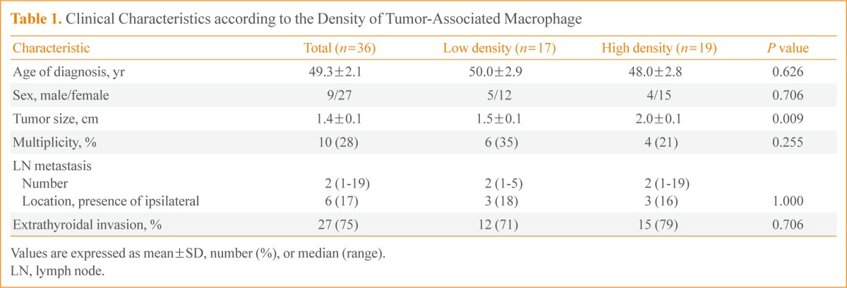
- The TAM densities (CD68+ cells) varied from 5% to 70%, in all tumor areas, while few cells were stained in adjacent normal tissues. TAMs were identified as CD68+ cells with thin, elongated cytoplasmic extensions that formed a canopy structure over tumor cells. Comparing clinicopathologic characteristics between tumors with low (<25%) and high (25% to 70%) TAM densities, primary tumors were larger in the high density group than in the low density group (2.0±0.1 vs. 1.5±0.1; P=0.009).
-
Conclusion
- TAMs were identified in primary PTC tumors with LN metastasis and higher TAM densities were related to larger tumor sizes, suggesting a tumorigenic role of TAMs in human PTCs.
- Macrophages are one of the components of bone marrow cells and play roles in innate immunity. Classically, they circulate in the blood and migrate into the inflammatory tissues such as infectious diseases in response to specific, endogenous immune signal [1,2]. Recently their alternative roles in anti-inflammation, cell clearance and tissue regenerations have also been established especially in inflammatory metabolic disorders including obesity and diabetes [1,2]. Therefore, macrophages have been categorized into two subtypes-M1 and M2-depending on their distinctive roles. M1 macrophages play proinflammatory functions in response to TH1 cytokines and M2 macrophages play anti-inflammatory and imunosuppressive activities in response to Th2 cytokines [3]. However, macrophages are not fixed to a single M1 or M2 subtype but suspected to rapidly differentiate and redifferentiate in various microenvironments.
- Tumor-associated macrophages (TAMs), macrophages existing tumor microenvironment, were first described in the early 1980s [4,5]. Till now, TAMs have been found to play dual functions, both positively or negatively affect tumor growth through interactions with the microenvironment, and these actions are tissue specific [6,7]. Several clinical studies showed that high density TAMs were present in more advanced stages of cancer with a worse prognosis in breast [8,9], lung [10,11], and bladder cancer [12,13]. Although a high TAM density has been reported in poorly differentiated papillary thyroid carcinoma (PTC) or anaplastic thyroid cancer [14], the distribution and function of TAMs in the most common PTC have not been studied yet. In this study, we have demonstrated the existence of TAMs in PTC tissue with lymph node (LN) metastasis and also used expression distribution and morphological properties to investigate the relationship between TAMs and the frequency of extrathyroidal invasion in PTC.
INTRODUCTION
- Study subjects
- The study was conducted from March to May 2012 at Seoul National University Hospital. The primary PTC lesion and surrounding normal tissues were collected from 36 consecutive patients with a previous thyroidectomy and central region LN dissection followed by a pathological diagnosis of PTC with LN metastasis. This diagnosis was defined as having a metastasized lesion in at least one LN from the histological examination of 1 to 37 LNs obtained from a central region LN dissection. All participating patients signed the informed consent form after receiving a comprehensive explanation of the study. This study was conducted with the approval of the Institutional Review Board of Seoul National University Hospital (IRB approval No. H-0809-0970-258).
- Histologic examination
- All sample tissues were fixed in 10% neutral formalin solution for 24 hours and washed with distilled water for 20 minutes before histological observation. Tissues were dehydrated with ethyl alcohol, washed with xylene, embedded in paraffin, and sectioned into 3 µm sections. Sections were deparaffinized, using a standard process of anhydrous ethyl alcohol, washed, and stained with hematoxilin and eosin. The size of the primary cancer and presence of extrathyroidal invasion (report of other pathological findings) were measured by two or more pathologists.
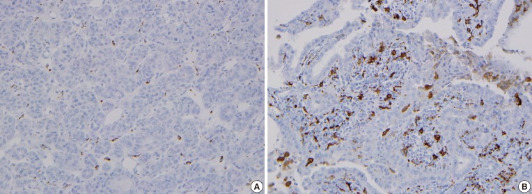
- For immunohistochemistry to demonstrate TAM expression, paraffin-embedded tissues were sectioned at 5 µm thick, deparaffinized, placed on a slide, dried at 37℃, and deparaffinized with xylene. They were then incubated with antihuman CD68 antibody PG-M1 (1:50, DakoCytomation, Carpinteria, CA, USA). Color staining was achieved with a DAKO EnVision+ system (DakoCytomation) by using a streptavidin biotin-horseradish peroxidase process and DAB (3,3'-diaminobenzi dine tetrahydrochloride) as a chromogen. Hematoxilin and eosin staining was used as a control. These sections were observed under an optical microscope. The proportion of CD68+ cells in each tumor on each slide was evaluated after staining. Based on a median value of 25%, weak positive (+) and strong positive (++) staining were defined as <25% and ≥25%, respectively (Fig. 1).
- Statistical analysis
- Patients' clinical characteristics are presented as mean±SEM. CD68 immunohistochemistry results were classified according to the pathological analysis along with a correlation analysis of other clinical data. Statistical significance is indicated with the P value, and a significant difference was considered when P values for both were less than 0.05. Statistical analysis was conducted by using SPSS version 17.0 (SPSS Inc., Chicago, IL, USA).
METHODS
- Clinical characteristics of study subjects
- The mean age of the 36 study subjects was 49.3±2.1 years old, and 27 subjects (75%) were female. The mean tumor size was 1.4±0.1 cm, and extrathyroidal invasion was observed in 27 subjects (75%). Ten subjects (28%) had multiple (two or three) tumors and eight of them had tumors in both lobes. LN metastases (median, 2; range, 1 to 19) were pathologically confirmed. Three metastases were found opposite the tumor, and three patients had bilateral LN metastases (Table 1).
- Characteristics of TAM staining in PTC
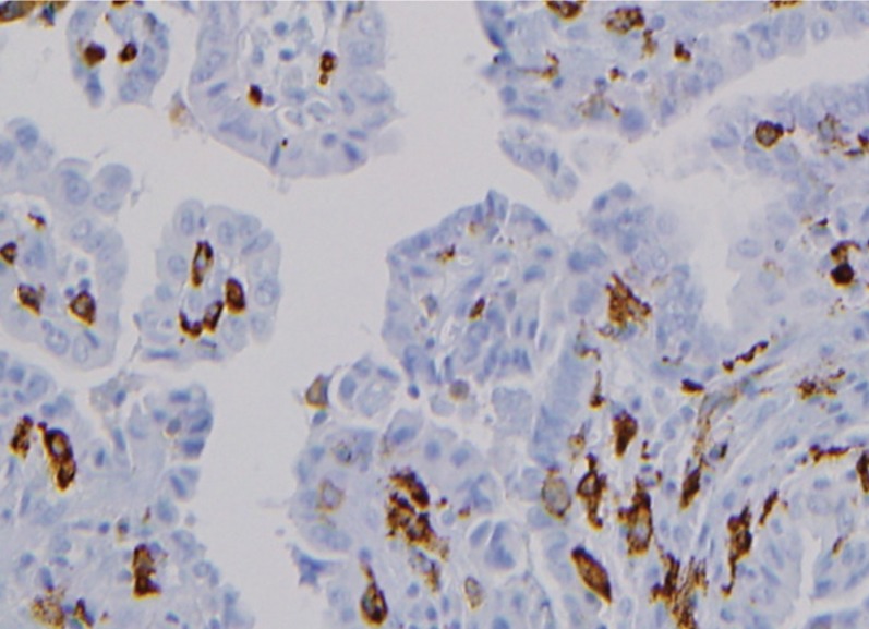
- The CD68 immunohistochemistry effectively stained macrophage cytoplasm and allowed us to observe TAMs in PTC. The TAM nuclei in PTC tissue were approximately 1/3 to 1/2 the size of the nuclei in surrounding tumor cells, and the cytoplasm was widely distributed, seeming to wrap around PTC cells. The cytoplasm of TAM and cancer cells appeared to be in close contact (Fig. 2). TAMs with extended nuclei were rare relative to the number of cancer cells due to the multidirectional expansion of TAM cytoplasm. The slide preparation may have affected the visibility of the cytoplasm, but not the layer containing the nucleus.
- Clinical characteristics for the distribution of TAM expression
- CD68+ cells were rare in normal thyroid tissue (Fig. 3A), but with the presence of TAMs, the CD68+ cells were present in PTC lesions. TAMs accounted 5% to 75% of all cancer cells, and were dispersed throughout the cancer tissues (Fig. 3B) or were grouped together in some lesions (Fig. 3C). This papillary arrangement of the thyroid cancer was maintained regardless of the presence of TAMs. There was no significant difference in clinical characteristics such as age at diagnosis, gender, size or number of primary tumor, or presence of extrathyroidal invasion according to the TAM expression pattern (diffuse or localized).
- Clinical characteristics by TAM density
- The patients' clinical characteristics were compared in tumors with low (17 patients) and high (19 patients) TAM density. TAM density was the proportion all cancer cells that were CD68+. The mean age at diagnosis and gender were not significantly different between the two groups, but the primary tumors were statistically larger in the group with high TAM density (1.5±0.1 vs. 2.0±0.1; P<0.01) (Table 1). The position and number of LN metastases was not analyzed statistically due to the small number of subject, but two patients with six or more LN metastases had TAM densities of 50% or higher. The number of tumors and presence of extrathyroidal invasion were similar between the two groups (Table 1).
RESULTS
- This study demonstrated the presence of TAM in a canopy structure over thyroid tumor cells in primary tumor tissues from 36 patients with PTC with LN metastasis. In addition, the primary tumors were significantly larger in the group with high TAM density. More LNs had metastases with a higher TAM density, although the difference was not statistically significant. Based on these findings, TAM density is considered to be related to PTC stage.
- Ryder et al. [14] demonstrated a higher TAM density in poorly differentiated PTC and anaplastic thyroid cancer than in well-differentiated PTC, by using CD68 and CD163 immunohistochemistry. Qing et al. [15] also reported a high TAM density in well-differentiated PTC, and the TAM density was significantly higher in tumors with TNM staging over stage III. The results from these studies demonstrated that a high TAM density is present in poorly differentiated PTC and is related to poor prognosis, which indicates TAMs may support PTC growth. However, the role of TAMs in thyroid cancer remains controversial. Fiumara et al. [16], using CD68 immunohistochemistry, found that TAM prevented metastasis in some patients. Of 121 patients with PTC, 15% had TAMs CD68+) that appeared to have a phagocytic function on cancer cells. These patients had significantly less blood vessel invasion and remote metastasis and largely accompanying invasions of lymphocytes and dendritic cells, than patients without an CD68+ cells. These results suggest that these cells may activate the host immune response to cancer cells. However, in our study, 56% of patients had TAMs without evidence of phagocytosis, and their clinical characteristics were similar to those without TAMs.
- This study investigated TAM morphologies and evaluated related clinical characteristics in PTC tissue with relatively advanced LN metastasis. Interestingly, CD68+ TAM cells observed in this study appeared to be mostly cytoplasm forming a canopy structure that projected over cancer cells. These canopy structures formed over the interior and exterior papillary projections in PTC cells that normally form papillary structures. These morphological characteristics are a standard of the TAM definition and differ from the rounded amoeba shape of inflammatory response cells, which are generally referred to as M1 macrophages [17]. The morphological characteristics of TAMs support cancer cell growth by forming a structural network between cancer cells and the extracellular matrix and are considered to support endothelial cells in forming blood vessels [18,19]. Caillou et al. [20] described the morphological characteristics of TAMs and their adjacency between cells, and these morphological characteristics suggest that TAMs facilitate cancer cell growth and metastasis. In this study, however, the morphological characteristics of thyroid cancer tissue were maintained regardless of the presence of TAMs, hence we suspect that TAMs do not have a huge effect on cancer development [20].
- Interestingly, despite of small number of patients, when we classified patients according to TAM density, the primary tumors were larger in the high TAM density group. Hence, we hypothesize that TAMs inhibit tumor growth in advanced PTC. These results differ from previous findings, where Qing et al. [15] compared the micropapillary cancer smaller than 1 cm and papillary cancer of typical size (1 cm or larger) and found no difference in TAM density. Apart from the etiological interpretation, TAM can be interpreted to be involved in cancer invasiveness after mobilization followed by the cancer growth. Actually, TAM has been related to metastasis in various cancer types [12].
- The study has a limitation of not including patients without LN metastasis. This study on TAMs reported that TAMs are rare in PTC, and especially they have only been reported in poorly differentiated cancer or anaplastic thyroid cancer. Therefore, this study only included the subjects with LN metastasis in higher stages of papillary cancer. A further study with more subjects including patients without LN metastasis is warranted.
- Studies on cancer treatments targeting TAM have increased since the first report that TAM facilitates cancer growth and metastasis and offers indicates a poor prognosis. Popular treatment strategies that have been investigated involve cytokines such as colony stimulating factor (CSF)-1 [21], which mobilizes TAM to the tumor; interleukin (IL)-10 [21] and IL-4 [22], which facilitate a cancer's affinity for TAM through macrophage differentiation into the M1 and M2 subtypes; and CCR2 [23,24], which facilitates TAM in cancer metastasis. Interestingly, a recent animal study was published that suggests that these strategies might be feasible in treating thyroid cancer [25]. Ryder et al. [25] demonstrated PTC inhibition in a BRAF-mutant, transformed mice model by inhibiting CSF-1 gene expression through genetic modification or use of a CSF-1/CSF-1 receptor signal pathway inhibitor.
- Studies on TAM in thyroid cancer are underway, but clinical data and studies on the mechanisms of TAM action are lacking. Thyroid cancer has a relatively favorable prognosis because its growth rate is slow. Therefore, general cancer pathophysiology does not apply to the role of TAMs. Currently the role of TAMs is only known in relatively invasive breast, lung, and pancreatic cancers, but the results of this study indicate that TAMs also exist in PTC, which is generally well differentiated with a favorable prognosis. Their role in cancer development is exciting; especially, the in light of the animal study targeting TAM that found possible inhibition of PTC. Expectations are high for treating patients with thyroid cancer since the BRAF mutation rate is higher among the Korean population than other ethnic groups. Additional study on the relationship between the BRAF mutation and TAM in Korean patients is warranted.
- This study was conducted with relatively few patients, but it demonstrated the presence of TAMs in clinically common PTC patients, and the presence of a high TAM density in larger primary cancers. However patients without LN metastasis were not included in the study or the comparative analysis. Another limitation arose from not evaluating the characteristics of TAMs in metastasized LNs. Also, there is a fundamental controversy over whether CD68, the TAM marker, is appropriate for the staining M2 macrophages, so the more specific CD 163 should be stained in future analyses.
- As a result of immunohistochemistry by using anti-CD68 antibody in PTC tissue with LN metastasis, TAM was present in all tissues with an extended cytoplasm forming a canopy shape over cancer cells. The density varied from 5% to 70% according to the tissue type. In normal thyroid tissue from the same patients, CD68+ cells were rare. When tumors were classified by TAM density, the primary tumors were statistically significantly larger in the group with a high TAM density. Therefore, TAM is suggested to stimulate cancer growth in PTC tissue with LN metastasis.
DISCUSSION
-
Acknowledgements
- This work was supported by a grant from the Next-Generation BioGreen 21 Program (No. PJ00954003), Rural Development Administration, Republic of Korea.
ACKNOWLEDGMENTS
- 1. Gordon S. Alternative activation of macrophages. Nat Rev Immunol 2003;3:23–35. ArticlePubMedPDF
- 2. Mosser DM. The many faces of macrophage activation. J Leukoc Biol 2003;73:209–212. ArticlePubMed
- 3. Mantovani A, Sozzani S, Locati M, Allavena P, Sica A. Macrophage polarization: tumor-associated macrophages as a paradigm for polarized M2 mononuclear phagocytes. Trends Immunol 2002;23:549–555. ArticlePubMed
- 4. Ladusch M, Schaffner H, Ullmann L, Littmann M, Reimann S, Gindl P, Ambrosius H. Pre- and postoperative reactivity of breast cancer patients to tumor associated antigens and HEP in the macrophage electrophoresis mobility (MEM) test. Arch Geschwulstforsch 1982;52:469–478. PubMed
- 5. Neumeister B, Hambsch K, Storch H. Macrophage adherence inhibition test (MAI) in Wistar rats bearing Jensen tumors. I. MAI after incubation with tumor-associated antigens. Arch Geschwulstforsch 1983;53:521–528. PubMed
- 6. Bingle L, Brown NJ, Lewis CE. The role of tumour-associated macrophages in tumour progression: implications for new anticancer therapies. J Pathol 2002;196:254–265. ArticlePubMed
- 7. Heusinkveld M, van der Burg SH. Identification and manipulation of tumor associated macrophages in human cancers. J Transl Med 2011;9:216ArticlePubMedPMCPDF
- 8. Tsutsui S, Yasuda K, Suzuki K, Tahara K, Higashi H, Era S. Macrophage infiltration and its prognostic implications in breast cancer: the relationship with VEGF expression and microvessel density. Oncol Rep 2005;14:425–431. ArticlePubMed
- 9. Campbell MJ, Tonlaar NY, Garwood ER, Huo D, Moore DH, Khramtsov AI, Au A, Baehner F, Chen Y, Malaka DO, Lin A, Adeyanju OO, Li S, Gong C, McGrath M, Olopade OI, Esserman LJ. Proliferating macrophages associated with high grade, hormone receptor negative breast cancer and poor clinical outcome. Breast Cancer Res Treat 2011;128:703–711. ArticlePubMedPDF
- 10. Hirayama S, Ishii G, Nagai K, Ono S, Kojima M, Yamauchi C, Aokage K, Hishida T, Yoshida J, Suzuki K, Ochiai A. Prognostic impact of CD204-positive macrophages in lung squamous cell carcinoma: possible contribution of Cd204-positive macrophages to the tumor-promoting microenvironment. J Thorac Oncol 2012;7:1790–1797. ArticlePubMed
- 11. Sato S, Hanibuchi M, Kuramoto T, Yamamori N, Goto H, Ogawa H, Mitsuhashi A, Van TT, Kakiuchi S, Akiyama S, Nishioka Y, Sone S. Macrophage stimulating protein promotes liver metastases of small cell lung cancer cells by affecting the organ microenvironment. Clin Exp Metastasis 2013;30:333–344. ArticlePubMedPDF
- 12. Zhang QW, Liu L, Gong CY, Shi HS, Zeng YH, Wang XZ, Zhao YW, Wei YQ. Prognostic significance of tumor-associated macrophages in solid tumor: a meta-analysis of the literature. PLoS One 2012;7:e50946ArticlePubMedPMC
- 13. Takayama H, Nishimura K, Tsujimura A, Nakai Y, Nakayama M, Aozasa K, Okuyama A, Nonomura N. Increased infiltration of tumor associated macrophages is associated with poor prognosis of bladder carcinoma in situ after intravesical bacillus Calmette-Guerin instillation. J Urol 2009;181:1894–1900. ArticlePubMed
- 14. Ryder M, Ghossein RA, Ricarte-Filho JC, Knauf JA, Fagin JA. Increased density of tumor-associated macrophages is associated with decreased survival in advanced thyroid cancer. Endocr Relat Cancer 2008;15:1069–1074. ArticlePubMedPMC
- 15. Qing W, Fang WY, Ye L, Shen LY, Zhang XF, Fei XC, Chen X, Wang WQ, Li XY, Xiao JC, Ning G. Density of tumor-associated macrophages correlates with lymph node metastasis in papillary thyroid carcinoma. Thyroid 2012;22:905–910. ArticlePubMedPMC
- 16. Fiumara A, Belfiore A, Russo G, Salomone E, Santonocito GM, Ippolito O, Vigneri R, Gangemi P. In situ evidence of neoplastic cell phagocytosis by macrophages in papillary thyroid cancer. J Clin Endocrinol Metab 1997;82:1615–1620. ArticlePubMed
- 17. Lawrence T, Natoli G. Transcriptional regulation of macrophage polarization: enabling diversity with identity. Nat Rev Immunol 2011;11:750–761. ArticlePubMedPDF
- 18. Lewis CE, Pollard JW. Distinct role of macrophages in different tumor microenvironments. Cancer Res 2006;66:605–612. ArticlePubMed
- 19. Mosser DM, Edwards JP. Exploring the full spectrum of macrophage activation. Nat Rev Immunol 2008;8:958–969. ArticlePubMedPMCPDF
- 20. Caillou B, Talbot M, Weyemi U, Pioche-Durieu C, Al Ghuzlan A, Bidart JM, Chouaib S, Schlumberger M, Dupuy C. Tumor-associated macrophages (TAMs) form an interconnected cellular supportive network in anaplastic thyroid carcinoma. PLoS One 2011;6:e22567ArticlePubMedPMC
- 21. Verreck FA, de Boer T, Langenberg DM, Hoeve MA, Kramer M, Vaisberg E, Kastelein R, Kolk A, de Waal-Malefyt R, Ottenhoff TH. Human IL-23-producing type 1 macrophages promote but IL-10-producing type 2 macrophages subvert immunity to (myco)bacteria. Proc Natl Acad Sci U S A 2004;101:4560–4565. ArticlePubMedPMC
- 22. Stein M, Keshav S, Harris N, Gordon S. Interleukin 4 potently enhances murine macrophage mannose receptor activity: a marker of alternative immunologic macrophage activation. J Exp Med 1992;176:287–292. ArticlePubMedPMCPDF
- 23. Penton-Rol G, Cota M, Polentarutti N, Luini W, Bernasconi S, Borsatti A, Sica A, LaRosa GJ, Sozzani S, Poli G, Mantovani A. Up-regulation of CCR2 chemokine receptor expression and increased susceptibility to the multitropic HIV strain 89.6 in monocytes exposed to glucocorticoid hormones. J Immunol 1999;163:3524–3529. ArticlePubMedPDF
- 24. Geissmann F, Jung S, Littman DR. Blood monocytes consist of two principal subsets with distinct migratory properties. Immunity 2003;19:71–82. ArticlePubMed
- 25. Ryder M, Gild M, Hohl TM, Pamer E, Knauf J, Ghossein R, Joyce JA, Fagin JA. Genetic and pharmacological targeting of CSF-1/CSF-1R inhibits tumor-associated macrophages and impairs BRAF-induced thyroid cancer progression. PLoS One 2013;8:e54302ArticlePubMedPMC
References


Figure & Data
References
Citations

- Single-cell RNA-seq reveals MIF−(CD74 + CXCR4) dependent inhibition of macrophages in metastatic papillary thyroid carcinoma
Wei Chen, Xinnian Yu, Huixin Li, Shenglong Yuan, Yuqi Fu, Huanhuan Hu, Fangzhou Liu, Yuan Zhang, Shanliang Zhong
Oral Oncology.2024; 148: 106654. CrossRef - H2AFX might be a prognostic biomarker for hepatocellular carcinoma
Hailiang Hu, Tao Zhong, Suwei Jiang
Cancer Reports.2023;[Epub] CrossRef - The Tumor Microenvironment and the Estrogen Loop in Thyroid Cancer
Nerina Denaro, Rebecca Romanò, Salvatore Alfieri, Alessia Dolci, Lisa Licitra, Imperia Nuzzolese, Michele Ghidini, Claudia Bareggi, Valentina Bertaglia, Cinzia Solinas, Ornella Garrone
Cancers.2023; 15(9): 2458. CrossRef - New Insights into Immune Cells and Immunotherapy for Thyroid Cancer
Yujia Tao, Peng Li, Chao Feng, Yuan Cao
Immunological Investigations.2023; 52(8): 1039. CrossRef - Roles and new Insights of Macrophages in the Tumor Microenvironment of Thyroid Cancer
Qi Liu, Wei Sun, Hao Zhang
Frontiers in Pharmacology.2022;[Epub] CrossRef - Construction of a Tumor Immune Microenvironment-Related Prognostic Model in BRAF-Mutated Papillary Thyroid Cancer
Yuxiao Xia, Xue Jiang, Yuan Huang, Qian Liu, Yin Huang, Bo Zhang, Zhanjun Mei, Dongkun Xu, Yuhong Shi, Wenling Tu
Frontiers in Endocrinology.2022;[Epub] CrossRef - Identification and Validation of Prognostic Related Hallmark ATP-Binding Cassette Transporters Associated With Immune Cell Infiltration Patterns in Thyroid Carcinoma
Lidong Wang, Xiaodan Sun, Jingni He, Zhen Liu
Frontiers in Oncology.2022;[Epub] CrossRef - Unraveling the Effects of Carotenoids Accumulation in Human Papillary Thyroid Carcinoma
Alessandra di Masi, Rosario Luigi Sessa, Ylenia Cerrato, Gianni Pastore, Barbara Guantario, Roberto Ambra, Michael Di Gioacchino, Armida Sodo, Martina Verri, Pierfilippo Crucitti, Filippo Longo, Anda Mihaela Naciu, Andrea Palermo, Chiara Taffon, Filippo A
Antioxidants.2022; 11(8): 1463. CrossRef - What is the status of immunotherapy in thyroid neoplasms?
Alejandro Garcia-Alvarez, Jorge Hernando, Ana Carmona-Alonso, Jaume Capdevila
Frontiers in Endocrinology.2022;[Epub] CrossRef - Matrix Metalloproteinase 9 Expression by Immunohistochemistry in Intratumor Macrophages as Tumor-Associated Macrophage Marker of Papillary Thyroid Carcinoma
Ni Wayan Armerinayanti, Samuel Widodo, Desak Putu Oki Lestari
Biomedical and Pharmacology Journal.2022; 15(3): 1671. CrossRef - Cell Component and Function of Tumor Microenvironment in Thyroid Cancer
Eunah Shin, Ja Seung Koo
International Journal of Molecular Sciences.2022; 23(20): 12578. CrossRef - Potential diagnostic of lymph node metastasis and prognostic values of TM4SFs in papillary thyroid carcinoma patients
Kun Wang, Haomin Li, Junyu Zhao, Jinming Yao, Yiran Lu, Jianjun Dong, Jie Bai, Lin Liao
Frontiers in Cell and Developmental Biology.2022;[Epub] CrossRef - Thyroid Cancer Stem-Like Cells: From Microenvironmental Niches to Therapeutic Strategies
Elisa Stellaria Grassi, Viola Ghiandai, Luca Persani
Journal of Clinical Medicine.2021; 10(7): 1455. CrossRef - Immune Landscape of Thyroid Cancers: New Insights
Elisa Menicali, Martina Guzzetti, Silvia Morelli, Sonia Moretti, Efisio Puxeddu
Frontiers in Endocrinology.2021;[Epub] CrossRef - FOXE1-Dependent Regulation of Macrophage Chemotaxis by Thyroid Cells In Vitro and In Vivo
Sara Credendino, Marta De Menna, Irene Cantone, Carmen Moccia, Matteo Esposito, Luigi Di Guida, Mario De Felice, Gabriella De Vita
International Journal of Molecular Sciences.2021; 22(14): 7666. CrossRef - Senescent Thyrocytes, Similarly to Thyroid Tumor Cells, Elicit M2-like Macrophage Polarization In Vivo
Mara Mazzoni, Giuseppe Mauro, Lucia Minoli, Loredana Cleris, Maria Chiara Anania, Tiziana Di Marco, Emanuela Minna, Sonia Pagliardini, Maria Grazia Rizzetti, Giacomo Manenti, Maria Grazia Borrello, Eugenio Scanziani, Angela Greco
Biology.2021; 10(10): 985. CrossRef - Characterization of the Immune Cell Infiltration Landscape of Thyroid Cancer for Improved Immunotherapy
Jing Gong, Bo Jin, Liang Shang, Ning Liu
Frontiers in Molecular Biosciences.2021;[Epub] CrossRef - The Thyroid Tumor Microenvironment: Potential Targets for Therapeutic Intervention and Prognostication
Laura MacDonald, Jonathan Jenkins, Grace Purvis, Joshua Lee, Aime T. Franco
Hormones and Cancer.2020; 11(5-6): 205. CrossRef - Cancer Stem Cells in Thyroid Tumors: From the Origin to Metastasis
Veronica Veschi, Francesco Verona, Melania Lo Iacono, Caterina D'Accardo, Gaetana Porcelli, Alice Turdo, Miriam Gaggianesi, Stefano Forte, Dario Giuffrida, Lorenzo Memeo, Matilde Todaro
Frontiers in Endocrinology.2020;[Epub] CrossRef - The Immune Landscape of Thyroid Cancer in the Context of Immune Checkpoint Inhibition
Gilda Varricchi, Stefania Loffredo, Giancarlo Marone, Luca Modestino, Poupak Fallahi, Silvia Martina Ferrari, Amato de Paulis, Alessandro Antonelli, Maria Rosaria Galdiero
International Journal of Molecular Sciences.2019; 20(16): 3934. CrossRef - Impact of tumor‐associated macrophages and BRAFV600E mutation on clinical outcomes in patients with various thyroid cancers
Jae Won Cho, Won Woong Kim, Yu‐mi Lee, Min Ji Jeon, Won Gu Kim, Dong Eun Song, Yangsoon Park, Ki‐Wook Chung, Suck Joon Hong, Tae‐Yon Sung
Head & Neck.2019; 41(3): 686. CrossRef - Senescent thyrocytes and thyroid tumor cells induce M2-like macrophage polarization of human monocytes via a PGE2-dependent mechanism
Mara Mazzoni, Giuseppe Mauro, Marco Erreni, Paola Romeo, Emanuela Minna, Maria Grazia Vizioli, Cristina Belgiovine, Maria Grazia Rizzetti, Sonia Pagliardini, Roberta Avigni, Maria Chiara Anania, Paola Allavena, Maria Grazia Borrello, Angela Greco
Journal of Experimental & Clinical Cancer Research.2019;[Epub] CrossRef - Immune and Inflammatory Cells in Thyroid Cancer Microenvironment
Ferrari, Fallahi, Galdiero, Ruffilli, Elia, Ragusa, Paparo, Patrizio, Mazzi, Varricchi, Marone, Antonelli
International Journal of Molecular Sciences.2019; 20(18): 4413. CrossRef - Aberrant Thyroid-Stimulating Hormone Receptor Signaling Increases VEGF-A and CXCL8 Secretion of Thyroid Cancer Cells, Contributing to Angiogenesis and Tumor Growth
Young Shin Song, Min Joo Kim, Hyun Jin Sun, Hwan Hee Kim, Hyo Shik Shin, Young A. Kim, Byung-Chul Oh, Sun Wook Cho, Young Joo Park
Clinical Cancer Research.2019; 25(1): 414. CrossRef - Novel targeted therapies and immunotherapy for advanced thyroid cancers
George E. Naoum, Michael Morkos, Brian Kim, Waleed Arafat
Molecular Cancer.2018;[Epub] CrossRef - Potential involvement of neutrophils in human thyroid cancer
Maria Rosaria Galdiero, Gilda Varricchi, Stefania Loffredo, Claudio Bellevicine, Tiziana Lansione, Anne Lise Ferrara, Raffaella Iannone, Sarah di Somma, Francesco Borriello, Eduardo Clery, Maria Triassi, Giancarlo Troncone, Gianni Marone, Fabrizio Mattei
PLOS ONE.2018; 13(6): e0199740. CrossRef - Bleomycin inhibits proliferation and induces apoptosis in TPC‑1 cells through reversing M2‑macrophages polarization
Hongwei Liu, Huilei Dong, Li Jiang, Zhendong Li, Xiande Ma
Oncology Letters.2018;[Epub] CrossRef - Pathological processes and therapeutic advances in radioiodide refractory thyroid cancer
Marika H Tesselaar, Johannes W Smit, James Nagarajah, Romana T Netea-Maier, Theo S Plantinga
Journal of Molecular Endocrinology.2017; 59(4): R141. CrossRef - The immune network in thyroid cancer
Maria Rosaria Galdiero, Gilda Varricchi, Gianni Marone
OncoImmunology.2016; 5(6): e1168556. CrossRef - Macrophage Densities Correlated with CXC Chemokine Receptor 4 Expression and Related with Poor Survival in Anaplastic Thyroid Cancer
Dae In Kim, Eunyoung Kim, Young A Kim, Sun Wook Cho, Jung Ah Lim, Young Joo Park
Endocrinology and Metabolism.2016; 31(3): 469. CrossRef - CXCL16 signaling mediated macrophage effects on tumor invasion of papillary thyroid carcinoma
Sun Wook Cho, Young A Kim, Hyun Jin Sun, Ye An Kim, Byung-Chul Oh, Ka Hee Yi, Do Joon Park, Young Joo Park
Endocrine-Related Cancer.2016; 23(2): 113. CrossRef - Role of pulmonary macrophages in initiation of lung metastasis in anaplastic thyroid cancer
Xiu Juan Li, Prakash Gangadaran, Senthilkumar Kalimuthu, Ji Min Oh, Liya Zhu, Shin Young Jeong, Sang‐Woo Lee, Jaetae Lee, Byeong‐Cheol Ahn
International Journal of Cancer.2016; 139(11): 2583. CrossRef - The Expression and Relationship of CD68-Tumor-Associated Macrophages and Microvascular Density With the Prognosis of Patients With Laryngeal Squamous Cell Carcinoma
Shujun Sun, Xinliang Pan, Limin Zhao, Jianming Zhou, Hongzeng Wang, Yonghong Sun
Clinical and Experimental Otorhinolaryngology.2016; 9(3): 270. CrossRef - Cancers with Higher Density of Tumor-Associated Macrophages Were Associated with Poor Survival Rates
Kyong Yeun Jung, Sun Wook Cho, Young A Kim, Daein Kim, Byung-Chul Oh, Do Joon Park, Young Joo Park
Journal of Pathology and Translational Medicine.2015; 49(4): 318. CrossRef - Association of the Preoperative Neutrophil-to-ymphocyte Count Ratio and Platelet-to-Lymphocyte Count Ratio with Clinicopathological Characteristics in Patients with Papillary Thyroid Cancer
Sang Mi Kim, Eun Heui Kim, Bo Hyun Kim, Jong Ho Kim, Su Bin Park, Yoon Jeong Nam, Kang Hee Ahn, Min Young Oh, Won Jin Kim, Yun Kyung Jeon, Sang Soo Kim, Yong Ki Kim, In Ju Kim
Endocrinology and Metabolism.2015; 30(4): 494. CrossRef - Quantitative Changes in Tumor-Associated M2 Macrophages Characterize Cholangiocarcinoma and their Association with Metastasis
Malinee Thanee, Watcharin Loilome, Anchalee Techasen, Nisana Namwat, Thidarut Boonmars, Chawalit Pairojkul, Puangrat Yongvanit
Asian Pacific Journal of Cancer Prevention.2015; 16(7): 3043. CrossRef - Tumor-Associated Macrophage (TAM) and Angiogenesis in Human Colon Carcinoma
Manal A. Badawi, Dalia M. Abouelfadl, Sonia L. El-Sharkawy, Wafaa E. Abd El-Aal, Naglaa F. Abbas
Open Access Macedonian Journal of Medical Sciences.2015; 3(2): 209. CrossRef - Brief Review of Articles in 'Endocrinology and Metabolism' in 2013
Won-Young Lee
Endocrinology and Metabolism.2014; 29(3): 251. CrossRef - miRNA Expression in Anaplastic Thyroid Carcinomas
Aline Hébrant, Sébastien Floor, Manuel Saiselet, Aline Antoniou, Alice Desbuleux, Bérengère Snyers, Caroline La, Nicolas de Saint Aubain, Emmanuelle Leteurtre, Guy Andry, Carine Maenhaut, Raul M. Luque
PLoS ONE.2014; 9(8): e103871. CrossRef - The Expression of Tumor-Associated Macrophages in Papillary Thyroid Carcinoma
Bo Hyun Kim
Endocrinology and Metabolism.2013; 28(3): 178. CrossRef

 KES
KES


 PubReader
 Cite
Cite


