
Articles
- Page Path
- HOME > Endocrinol Metab > Volume 33(2); 2018 > Article
-
Review ArticleManagement of Low-Risk Papillary Thyroid Cancer
-
Nicole M. Iñiguez-Ariza1,2
 , Juan P. Brito2,3
 -
Endocrinology and Metabolism 2018;33(2):185-194.
DOI: https://doi.org/10.3803/EnM.2018.33.2.185
Published online: June 21, 2018
1Department of Endocrinology and Metabolism, Instituto Nacional de Ciencias Médicas y Nutrición Salvador Zubirán, Mexico City, Mexico.
2Division of Endocrinology, Diabetes, Metabolism, and Nutrition, Mayo Clinic, Rochester, MN, USA.
3Knowledge and Evaluation Research Unit, Mayo Clinic, Rochester, MN, USA.
- Corresponding author: Juan P. Brito. Division of Endocrinology, Diabetes, Metabolism, and Nutrition, and Knowledge and Evaluation Research Unit, Mayo Clinic, 200 First St. SW, Rochester, MN 55905, USA. Tel: +1-507-284-2463, Fax: +1-507-284-5745, brito.juan@mayo.edu
• Received: May 4, 2018 • Revised: May 14, 2018 • Accepted: May 21, 2018
Copyright © 2018 Korean Endocrine Society
This is an Open Access article distributed under the terms of the Creative Commons Attribution Non-Commercial License (http://creativecommons.org/licenses/by-nc/4.0/) which permits unrestricted non-commercial use, distribution, and reproduction in any medium, provided the original work is properly cited.
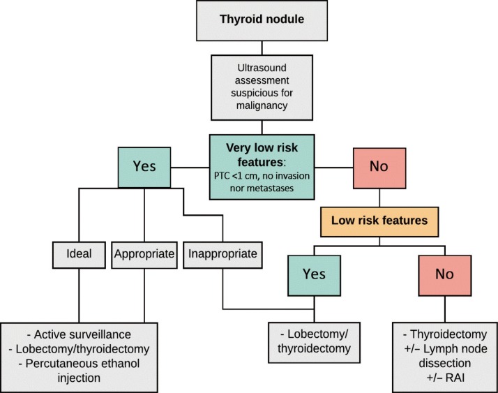
Figure & Data
References
Citations
Citations to this article as recorded by 
- Risk of malignancy in Thy3 thyroid nodules
Emad Mofid Nassif Rezkallah, Ragai Sobhi Hanna, Wael Magdy Elsaify
Endocrine Regulations.2024; 58(1): 19. CrossRef - Adequacy of clinical guideline recommendations for patients with low-risk cancer managed with monitoring: systematic review
Kiana K. Collins, Claire Friedemann Smith, Tori Ford, Nia Roberts, Brian D. Nicholson, Jason L. Oke
Journal of Clinical Epidemiology.2024; 169: 111280. CrossRef - Integrated metabolic and genetic analysis reveals distinct features of human differentiated thyroid cancer
Eduardo Cararo Lopes, Akshada Sawant, Dirk Moore, Hua Ke, Fuqian Shi, Saurabh Laddha, Ying Chen, Anchal Sharma, Jake Naumann, Jessie Yanxiang Guo, Maria Gomez, Maria Ibrahim, Tracey L. Smith, Gregory M. Riedlinger, Edmund C. Lattime, Stanley Trooskin, Shr
Clinical and Translational Medicine.2023;[Epub] CrossRef - Clinical management of low-risk papillary thyroid microcarcinoma
Chie MASAKI, Kiminori SUGINO, Koichi ITO
Minerva Endocrinology.2022;[Epub] CrossRef - The circ_FAM53B-miR-183-5p-CCDC6 axis modulates the malignant behaviors of papillary thyroid carcinoma cells
Chong Zhang, Huxia Gu, Dingrong Liu, Fuyun Tong, Huijie Wei, Dan Zhou, Jing Fang, Xiaolu Dai, Haibo Tian
Molecular and Cellular Biochemistry.2022; 477(11): 2627. CrossRef - An Appraisal and Update of Fluorodeoxyglucose and Non-Fluorodeoxyglucose-PET Tracers in Thyroid and Non–Thyroid Endocrine Neoplasms
Aadil Adnan, Shobhana Raju, Rakesh Kumar, Sandip Basu
PET Clinics.2022; 17(3): 343. CrossRef - A Narrative Review of Preventive Central Lymph Node Dissection in Patients With Papillary Thyroid Cancer - A Necessity or an Excess
David D. Dolidze, Alexey V. Shabunin, Robert B. Mumladze, Arshak V. Vardanyan, Serghei D. Covantsev, Alexander M. Shulutko, Vasiliy I. Semikov, Khalid M. Isaev, Airazat M. Kazaryan
Frontiers in Oncology.2022;[Epub] CrossRef - Ethanol Ablation as a Treatment in a Low-Risk Follicular Thyroid Cancer: A Case Report
Juan Pesantez, Carla Lituma, Carla Valencia, Jose Prieto, Marco Cazorla
Cureus.2022;[Epub] CrossRef - The new T3b category has clinical significance? SEER‐based study
Jingzhe Xiang, Zhihong Wang, Wei Sun, Hao Zhang
Clinical Endocrinology.2021; 94(3): 449. CrossRef - Efficacy and Safety of Thermal Ablation for Solitary T1bN0M0 Papillary Thyroid Carcinoma: A Multicenter Study
Xiao-Jing Cao, Juan Liu, Ya-Lin Zhu, Lu Qi, Geng Liu, Hong-Ling Wang, Zhong-Hua Wang, Ying Zhou, Jun-Feng He, Jian-Qin Guo, Li-Li Shi, Mei Jian, Aini Shataer, Guo-Zhen Yan, Zhen-Long Zhao, Ying Wei, Li-Li Peng, Yan Li, Ying Che, Shu-Rong Wang, Ming-An Yu
The Journal of Clinical Endocrinology & Metabolism.2021; 106(2): e573. CrossRef - A Clinical Audit of Hemithyroidectomy for Differentiated Thyroid Cancer—Experience from a Tertiary Cancer Center
Nithyanand Chidambaranathan, Shivakumar Thiagarajan, Nandini Menon, Adhara Chakraborthy, Richa Vaish, Devendra Chaukar
Indian Journal of Surgery.2021; 83(6): 1444. CrossRef - Pros and cons of hemi‐thyroidectomy for low‐risk differentiated thyroid cancer
Alexander J. Papachristos, Anthony Glover, Mark S. Sywak, Stan B. Sidhu
ANZ Journal of Surgery.2021; 91(9): 1704. CrossRef - Lactate Dehydrogenase A as a Potential New Biomarker for Thyroid Cancer
Eun Jeong Ban, Daham Kim, Jin Kyong Kim, Sang-Wook Kang, Jandee Lee, Jong Ju Jeong, Kee-Hyun Nam, Woong Youn Chung, Kunhong Kim
Endocrinology and Metabolism.2021; 36(1): 96. CrossRef - Taraxasterol inhibits TGF-β1-induced epithelial-to-mesenchymal transition in papillary thyroid cancer cells through regulating the Wnt/β-catenin signaling
J Zhu, X Li, S Zhang, J Liu, X Yao, Q Zhao, B Kou, P Han, X Wang, Y Bai, Z Zheng, C Xu
Human & Experimental Toxicology.2021; 40(12_suppl): S87. CrossRef - Is Delayed Papillary Thyroid Cancer Surgery Associated with Higher Mortality Risk?
Eddy P. Lincango Naranjo, Juan P. Brito
Clinical Thyroidology.2021; 33(9): 400. CrossRef - MicroRNA-99a-3p/GRP94 axis affects metastatic progression of human papillary thyroid carcinoma by regulating ITGA2 expression and localization
Yun Gao, Yi Pan, Tingting Wang, Ying Yao, Wenbo Yuan, Xue Zhu, Ke Wang
Acta Biochimica et Biophysica Sinica.2021; 53(12): 1650. CrossRef - Microscopic Extrathyroidal Extension Results in Increased Rate of Tumor Recurrence and Is an Independent Predictor of Patient’s Outcome in Middle Eastern Papillary Thyroid Carcinoma
Sandeep Kumar Parvathareddy, Abdul K. Siraj, Zeeshan Qadri, Felisa DeVera, Khawar Siddiqui, Saif S. Al-Sobhi, Fouad Al-Dayel, Khawla S. Al-Kuraya
Frontiers in Oncology.2021;[Epub] CrossRef - New paradigms in the treatment of low-risk thyroid cancer
Firas Baidoun, Anas M. Saad, Omar Abdel-Rahman
Expert Review of Endocrinology & Metabolism.2020; 15(4): 251. CrossRef - The Impact of Transcription Factor Prospero Homeobox 1 on the Regulation of Thyroid Cancer Malignancy
Magdalena Rudzińska, Barbara Czarnocka
International Journal of Molecular Sciences.2020; 21(9): 3220. CrossRef - Modified risk stratification based on cervical lymph node metastases following lobectomy for papillary thyroid carcinoma
Eyun Song, Jonghwa Ahn, Dong Eun Song, Won Woong Kim, Min Ji Jeon, Tae‐Yon Sung, Tae Yong Kim, Ki Wook Chung, Won Bae Kim, Young Kee Shong, Suck Joon Hong, Yu‐Mi Lee, Won Gu Kim
Clinical Endocrinology.2020; 92(4): 358. CrossRef - Clinicopathological significance of the single nucleotide polymorphism, rs2853669 within the TERT promoter in papillary thyroid carcinoma
Tatsuya Hirokawa, Yuu Arimasu, Tomohiro Chiba, Masachika Fujiwara, Hiroshi Kamma
Pathology International.2020; 70(4): 217. CrossRef - Diagnostic accuracy and ability to reduce unnecessary FNAC: A comparison between four Thyroid Imaging Reporting Data System (TI-RADS) versions
Lingsze Tan, Ying Sern Tan, Suzet Tan
Clinical Imaging.2020; 65: 133. CrossRef - Amino Acid Transporters as Potential Therapeutic Targets in Thyroid Cancer
Keisuke Enomoto, Muneki Hotomi
Endocrinology and Metabolism.2020; 35(2): 227. CrossRef - FDG PET/CT versus somatostatin receptor PET/CT in TENIS syndrome: a systematic review and meta-analysis
Felipe Alves Mourato, Maria Amorim Almeida, Ana Emília Teixeira Brito, Aline Lopes Garcia Leal, Paulo Almeida Filho, Elba Etchebehere
Clinical and Translational Imaging.2020; 8(5): 365. CrossRef - Factors Associated with Health Behaviors in Thyroid Cancer Survivors
Junghyun Yoon, Boyoung Park
Journal of Cancer Prevention.2020; 25(3): 173. CrossRef - Patient-Derived Papillary Thyroid Cancer Organoids for Radioactive Iodine Refractory Screening
Luc H.J. Sondorp, Vivian M.L. Ogundipe, Andries H. Groen, Wendy Kelder, Annelies Kemper, Thera P. Links, Robert P. Coppes, Schelto Kruijff
Cancers.2020; 12(11): 3212. CrossRef - Hemithyroidectomy for Thyroid Cancer: A Review
Noor Addasi, Abbey Fingeret, Whitney Goldner
Medicina.2020; 56(11): 586. CrossRef - Clinical Outcomes after Early and Delayed Radioiodine Remnant Ablation in Patients with Low-Risk Papillary Thyroid Carcinoma: Propensity Score Matching Analysis
Jonghwa Ahn, Meihua Jin, Eyun Song, Min Ji Jeon, Tae Yong Kim, Jin-Sook Ryu, Won Bae Kim, Young Kee Shong, Ji Min Han, Won Gu Kim
Endocrinology and Metabolism.2020; 35(4): 830. CrossRef - Clinical Significance of Gross Invasion of Strap Muscles in Patients With 1- to 4-cm-Sized Papillary Thyroid Carcinoma Undergoing Lobectomy
Eyun Song, Won Woong Kim, Min Ji Jeon, Tae-Yon Sung, Dong Eun Song, Tae Yong Kim, Ki Wook Chung, Won Bae Kim, Young Kee Shong, Suck Joon Hong, Yu-Mi Lee, Won Gu Kim
Annals of Surgical Oncology.2019; 26(13): 4466. CrossRef - Lobectomy Is Feasible for 1–4 cm Papillary Thyroid Carcinomas: A 10-Year Propensity Score Matched-Pair Analysis on Recurrence
Eyun Song, Minkyu Han, Hye-Seon Oh, Won Woong Kim, Min Ji Jeon, Yu-Mi Lee, Tae Yong Kim, Ki Wook Chung, Won Bae Kim, Young Kee Shong, Suck Joon Hong, Tae-Yon Sung, Won Gu Kim
Thyroid.2019; 29(1): 64. CrossRef - Laser and radiofrequency ablations for benign and malignant thyroid tumors
Giovanni Mauri, Nicolò Gennaro, Min Kyoung Lee, Jung Hwan Baek
International Journal of Hyperthermia.2019; 36(2): 13. CrossRef - US-Guided Radiofrequency Ablation for Low-Risk Papillary Thyroid Microcarcinoma: Efficacy and Safety in a Large Population
Hyun Kyung Lim, Se Jin Cho, Jung Hwan Baek, Kang Dae Lee, Chang Woo Son, Jung Min Son, Seon Mi Baek
Korean Journal of Radiology.2019; 20(12): 1653. CrossRef - Utility of Early Postoperative Unstimulated Thyroglobulin in Influencing Decision Making in Patients with Papillary Thyroid Carcinoma
Alexandria D. McDow, Cynthia M. Shumway, Susan C. Pitt, David F. Schneider, Rebecca S. Sippel, Kristin L. Long
Annals of Surgical Oncology.2019; 26(12): 4002. CrossRef - Extent of Surgery for Low-Risk Differentiated Thyroid Cancer
Alexandria D. McDow, Susan C. Pitt
Surgical Clinics of North America.2019; 99(4): 599. CrossRef
 PubReader
 ePub Link
 Cite
Cite

