
Search
- Page Path
- HOME > Search
Review Article
- Hypothalamus and Pituitary Gland
- Independent Skeletal Actions of Pituitary Hormones
- Se-Min Kim, Farhath Sultana, Funda Korkmaz, Daria Lizneva, Tony Yuen, Mone Zaidi
- Endocrinol Metab. 2022;37(5):719-731. Published online September 28, 2022
- DOI: https://doi.org/10.3803/EnM.2022.1573
- 3,668 View
- 235 Download
- 4 Web of Science
- 4 Crossref
-
 Abstract
 PDF
PDF PubReader
  ePub
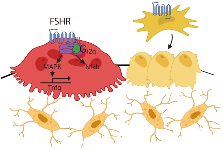
ePub - Over the past years, pituitary hormones and their receptors have been shown to have non-traditional actions that allow them to bypass the hypothalamus-pituitary-effector glands axis. Bone cells—osteoblasts and osteoclasts—express receptors for growth hormone, follicle stimulating hormone (FSH), thyroid stimulating hormone (TSH), adrenocorticotrophic hormone (ACTH), prolactin, oxytocin, and vasopressin. Independent skeletal actions of pituitary hormones on bone have been studied using genetically modified mice with haploinsufficiency and by activating or inactivating the receptors pharmacologically, without altering systemic effector hormone levels. On another front, the discovery of a TSH variant (TSH-βv) in immune cells in the bone marrow and skeletal action of FSHβ through tumor necrosis factor α provides new insights underscoring the integrated physiology of bone-immune-endocrine axis. Here we discuss the interaction of each pituitary hormone with bone and the potential it holds in understanding bone physiology and as a therapeutic target.
-
Citations
Citations to this article as recorded by
- New tools for bone health assessment in secreting pituitary adenomas
Meliha Melin Uygur, Stefano Frara, Luigi di Filippo, Andrea Giustina
Trends in Endocrinology & Metabolism.2023; 34(4): 231. CrossRef - A Causality between Thyroid Function and Bone Mineral Density in Childhood: Abnormal Thyrotropin May Be Another Pediatric Predictor of Bone Fragility
Dongjin Lee, Moon Ahn
Metabolites.2023; 13(3): 372. CrossRef - The mechanism of oxytocin and its receptors in regulating cells in bone metabolism
Liu Feixiang, Feng Yanchen, Li Xiang, Zhang Yunke, Miao Jinxin, Wang Jianru, Lin Zixuan
Frontiers in Pharmacology.2023;[Epub] CrossRef - To investigate the mechanism of Yiwei Decoction in the treatment of premature ovarian insufficiency-related osteoporosis using transcriptomics, network pharmacology and molecular docking techniques
Weisen Fan, Yan Meng, Jing Zhang, Muzhen Li, Yingjie Zhang, Xintian Qu, Xin Xiu
Scientific Reports.2023;[Epub] CrossRef
- New tools for bone health assessment in secreting pituitary adenomas

Original Article
- Endocrine Research
- Effects of Oxytocin on Cell Proliferation in a Corticotroph Adenoma Cell Line
- Jung Soo Lim, Young Woo Eom, Eun Soo Lee, Hyeong Ju Kwon, Ja-Young Kwon, Junjeong Choi, Choon Hee Chung, Young Suk Jo, Eun Jig Lee
- Endocrinol Metab. 2019;34(3):302-313. Published online September 26, 2019
- DOI: https://doi.org/10.3803/EnM.2019.34.3.302
- 5,010 View
- 74 Download
- 3 Web of Science
- 2 Crossref
-
Abstract
PDF
 Supplementary MaterialPubReader ePub
 Background Oxytocin (OXT) has been reported to act as a growth regulator in various tumor cells. However, there is a paucity of data on the influence of OXT on cell proliferation of corticotroph adenomas. This study aimed to examine whether OXT affects cell growth in pituitary tumor cell lines (AtT20 and GH3 cells) with a focus on corticotroph adenoma cells.
Methods Reverse transcription polymerase chain reaction and enzyme-linked immunosorbent assay were conducted with AtT20 cells to confirm the effects of OXT on hormonal activity; flow cytometry was used to assess changes in the cell cycle after OXT treatment. Moreover, the impact of OXT on proliferating cell nuclear antigen (PCNA), nuclear factor κB, and mitogen-activated protein kinase signaling pathway was analyzed by Western blot.
Results OXT treatment of 50 nM changed the gene expression of OXT receptor and pro-opiomelanocortin within a short time. In addition, OXT significantly reduced adrenocorticotropic hormone secretion within 1 hour. S and G2/M populations of AtT20 cells treated with OXT for 24 hours were significantly decreased compared to the control. Furthermore, OXT treatment decreased the protein levels of PCNA and phosphorylated extracellular-signal-regulated kinase (P-ERK) in AtT20 cells.
Conclusion Although the cytotoxic effect of OXT in AtT20 cells was not definite, OXT may blunt cell proliferation of corticotroph adenomas by altering the cell cycle or reducing PCNA and P-ERK levels. Further research is required to investigate the role of OXT as a potential therapeutic target in corticotroph adenomas.
-
Citations
Citations to this article as recorded by- Increased proliferation and neuronal fate in prairie vole brain progenitor cells cultured in vitro: effects by social exposure and sexual dimorphism
Daniela Ávila-González, Italo Romero-Morales, Lizette Caro, Alejandro Martínez-Juárez, Larry J. Young, Francisco Camacho-Barrios, Omar Martínez-Alarcón, Analía E. Castro, Raúl G. Paredes, Néstor F. Díaz, Wendy Portillo
Biology of Sex Differences.2023;[Epub] CrossRef - Anterior pituitary gland synthesises dopamine from l‐3,4‐dihydroxyphenylalanine (l‐dopa)
Santiago Jordi Orrillo, Nataly de Dios, Antonela Sofía Asad, Fernanda De Fino, Mercedes Imsen, Ana Clara Romero, Sandra Zárate, Jimena Ferraris, Daniel Pisera
Journal of Neuroendocrinology.2020;[Epub] CrossRef
- Increased proliferation and neuronal fate in prairie vole brain progenitor cells cultured in vitro: effects by social exposure and sexual dimorphism
Review Article
- Kisspeptin Regulation of Neuronal Activity throughout the Central Nervous System
- Xinhuai Liu, Allan E. Herbison
- Endocrinol Metab. 2016;31(2):193-205. Published online May 27, 2016
- DOI: https://doi.org/10.3803/EnM.2016.31.2.193
- 5,999 View
- 78 Download
- 38 Web of Science
- 37 Crossref
-
Abstract
PDFPubReader
Kisspeptin signaling at the gonadotropin-releasing hormone (GnRH) neuron is now relatively well characterized and established as being critical for the neural control of fertility. However, kisspeptin fibers and the kisspeptin receptor (KISS1R) are detected throughout the brain suggesting that kisspeptin is involved in regulating the activity of multiple neuronal circuits. We provide here a review of kisspeptin actions on neuronal populations throughout the brain including the magnocellular oxytocin and vasopressin neurons, and cells within the arcuate nucleus, hippocampus, and amygdala. The actions of kisspeptin in these brain regions are compared to its effects upon GnRH neurons. Two major themes arise from this analysis. First, it is apparent that kisspeptin signaling through KISS1R at the GnRH neuron is a unique, extremely potent form or neurotransmission whereas kisspeptin actions through KISS1R in other brain regions exhibit neuromodulatory actions typical of other neuropeptides. Second, it is becoming increasingly likely that kisspeptin acts as a neuromodulator not only through KISS1R but also through other RFamide receptors such as the neuropeptide FF receptors (NPFFRs). We suggest likely locations of kisspeptin signaling through NPFFRs but note that only limited tools are presently available for examining kisspeptin cross-signaling within the RFamide family of neuropeptides.
-
Citations
Citations to this article as recorded by- Role of the histone variant H2A.Z.1 in memory, transcription, and alternative splicing is mediated by lysine modification
Anas Reda, Luca A. Hategan, Timothy A. B. McLean, Samantha D. Creighton, Jian Qi Luo, Sean En Si Chen, Shan Hua, Stephen Winston, Isaiah Reeves, Aditya Padmanabhan, Tarkan A. Dahi, Firyal Ramzan, Mark A. Brimble, Patrick J. Murphy, Brandon J. Walters, Gil
Neuropsychopharmacology.2024;[Epub] CrossRef - System-wide mapping of peptide-GPCR interactions in C. elegans
Isabel Beets, Sven Zels, Elke Vandewyer, Jonas Demeulemeester, Jelle Caers, Esra Baytemur, Amy Courtney, Luca Golinelli, İlayda Hasakioğulları, William R. Schafer, Petra E. Vértes, Olivier Mirabeau, Liliane Schoofs
Cell Reports.2023; 42(9): 113058. CrossRef - Impose of KNDy/GnRH neural circuit in PCOS, ageing, cancer and Alzheimer’s disease: StAR actions in prevention of neuroendocrine dysfunction
Siva Prasad Panda, Adarsh Kesharwani, Gaurav Deep Singh, DSNBK Prasanth, Bhaskara Raju Vatchavai, P.V. Kamala Kumari, Sunil Kumar Panda, Sarada Prasanna Mallick
Ageing Research Reviews.2023; 92: 102086. CrossRef - Gallium-68-Labeled KISS1-54 Peptide for Mapping KISS1 Receptor via PET: Initial Evaluation in Human Tumor Cell Lines and in Tumor-Bearing Mice
Ina Israel, Gabriele Riehl, Elke Butt, Andreas K. Buck, Samuel Samnick
Pharmaceuticals.2023; 17(1): 44. CrossRef - Kisspeptin-13 prevented the electrophysiological alterations induced by amyloid-beta pathology in rat: Possible involvement of stromal interaction molecules and pCREB
Shima Ebrahimi Khonacha, Seyed Hamidreza Mirbehbahani, Mona Rahdar, Shima Davoudi, Mehdi Borjkhani, Fariba Khodagholi, Fereshteh Motamedi, Mahyar Janahmadi
Brain Research Bulletin.2022; 184: 13. CrossRef - Sexual Dimorphism in Kisspeptin Signaling
Eun Bee Lee, Iman Dilower, Courtney A. Marsh, Michael W. Wolfe, Saeed Masumi, Sameer Upadhyaya, Mohammad A. Karim Rumi
Cells.2022; 11(7): 1146. CrossRef - Estradiol and progesterone-induced lordosis behavior is modulated by both the Kisspeptin receptor and melanin-concentrating hormone in estradiol benzoate-primed rats
Oscar González-Flores, James G. Pfaus, Ailyn Luna-Hernández, Omar Montes-Narváez, Raymundo Domínguez-Ordóñez, Miriam B. Tecamachaltzi-Silvarán, Marcos García-Juárez
Hormones and Behavior.2022; 146: 105257. CrossRef - Targeting KNDy neurons to control GnRH pulses
Stephanie Constantin
Current Opinion in Pharmacology.2022; 67: 102316. CrossRef -
The ameliorative effects of
Lactobacillus coagulans
and
Lactobacillus casei
probiotics on CCl4‐induced testicular toxicity based on biochemical, histological and molecular analyses in rat
Zahra Keshtmand, Mohsen Akbaribazm, Yasin Bagheri, Reyhaneh Oliaei
Andrologia.2021;[Epub] CrossRef - Nitric oxide resets kisspeptin-excited GnRH neurons via PIP2 replenishment
Stephanie Constantin, Daniel Reynolds, Andrew Oh, Katherine Pizano, Susan Wray
Proceedings of the National Academy of Sciences.2021;[Epub] CrossRef - Expression analysis of neuropeptide FF receptors on neuroendocrine-related neurons in the rat brain using highly sensitive in situ hybridization
Shimpei Higo, Moeko Kanaya, Hitoshi Ozawa
Histochemistry and Cell Biology.2021; 155(4): 465. CrossRef - Timing the Juvenile-Adult Neurohormonal Transition: Functions and Evolution
Celia G. Barredo, Beatriz Gil-Marti, Derya Deveci, Nuria M. Romero, Francisco A. Martin
Frontiers in Endocrinology.2021;[Epub] CrossRef - Kisspeptin-54 attenuates oxidative stress and neuronal apoptosis in early brain injury after subarachnoid hemorrhage in rats via GPR54/ARRB2/AKT/GSK3β signaling pathway
Yi Huang, Yong Guo, Lei Huang, Yuanjian Fang, Dujuan Li, Rui Liu, Qin Lu, Reng Ren, Lihui Tang, Lifei Lian, Yongmei Hu, Jiping Tang, Gao Chen, John H. Zhang
Free Radical Biology and Medicine.2021; 171: 99. CrossRef - Conditioned place preference of kisspeptin-10
Ilia Yu. Tissen, Polina A. Chepik, Andrei A. Lebedev, Leila A. Magarramova, Eugenii R. Bychkov, Petr D. Shabanov
Reviews on Clinical Pharmacology and Drug Therapy.2021; 19(1): 47. CrossRef - Expression of type one cannabinoid receptor in different subpopulation of kisspeptin neurons and kisspeptin afferents to GnRH neurons in female mice
Tamás Wilheim, Krisztina Nagy, Mahendravarman Mohanraj, Kamil Ziarniak, Masahiko Watanabe, Joanna Sliwowska, Imre Kalló
Brain Structure and Function.2021; 226(7): 2387. CrossRef - Dopamine D2 Receptor Antagonist Alters the Testosterone Release and Kisspeptin/GPR54 Signaling in Food-Restricted Rats
Khatereh Nourmohammadi, Farrin Babaei-Balderlou, Seyyed Meysam Abtahi-Foroushani
Iranian Journal of Science and Technology, Transactions A: Science.2021; 45(6): 1879. CrossRef - Astrocytic Hydrogen Sulfide Regulates Supraoptic Cellular Activity in the Adaptive Response of Lactating Rats to Chronic Social Stress
Dongyang Li, Haitao Liu, Hongyang Wang, Shuwei Jia, Xiaoran Wang, Shuo Ling, Guichuan Chen, Xiaoyu Liu, Yu-Feng Wang
ASN Neuro.2021; 13: 175909142110430. CrossRef - Experimental characterization of a biosensor based on a tapered optical fiber for kisspeptin detection
K. González-León, G. Beltrán-Pérez, S. Muñoz-Aguirre, V. López -Gayou, J. Castillo-Mixcoatl, V. Alatriste, R. Delgado-Macuil
Applied Optics.2020; 59(13): D131. CrossRef - Cloning, characterisation and expression profile of kisspeptin1 and the kisspeptin1 receptor in the hypothalamic–pituitary–ovarian axis of Chinese alligator Alligator sinensis during the reproductive cycle
Ruidong Zhang, Haitao Nie, Shulong Duan, Peng Yan, Ali Izaz, Renping Wang, Yongkang Zhou, Xiaobing Wu
Reproduction, Fertility and Development.2020; 32(8): 792. CrossRef - Multiple failures in the lutenising hormone surge generating system in GABAB1KO female mice
Noelia P. Di Giorgio, Marianne Bizzozzero Hiriart, Pablo N. Surkin, Paula V. López, Nadia S. Bourguignon, Verónica B. Dorfman, Bernhard Bettler, Carlos Libertun, Victoria Lux‐Lantos
Journal of Neuroendocrinology.2019;[Epub] CrossRef - Vasopressin regulates hypothalamic GnRH synthesis: Histomorphological evidence in hypothalamus and biological effects in GT1-7 cells
Zhu Zhu, Xiaozhen Zhao, Feng Huang, Feng Wang, Wei Wang
Life Sciences.2019; 227: 166. CrossRef - The kisspeptin derivative kissorphin reduces the acquisition, expression, and reinstatement of ethanol-induced conditioned place preference in rats
Ewa Gibula-Tarlowska, Pawel Grochecki, Jerzy Silberring, Jolanta H. Kotlinska
Alcohol.2019; 81: 11. CrossRef - Unraveling the connection between GABA and kisspeptin in the control of reproduction
Noelia P Di Giorgio, Marianne Bizzozzero-Hiriart, Carlos Libertun, Victoria Lux-Lantos
Reproduction.2019; 157(6): R225. CrossRef - Region-specific changes in brain kisspeptin receptor expression during estrogen depletion and the estrous cycle
Saeko Ozaki, Shimpei Higo, Kinuyo Iwata, Hidehisa Saeki, Hitoshi Ozawa
Histochemistry and Cell Biology.2019; 152(1): 25. CrossRef - The long and the short of it – a perspective on peptidergic regulation of circuits and behaviour
Gáspár Jékely, Sarah Melzer, Isabel Beets, Ilona C. Grunwald Kadow, Joris Koene, Sara Haddad, Lindy Holden-Dye
Journal of Experimental Biology.2018;[Epub] CrossRef - Biological Significance of Kisspeptin–Kiss 1 Receptor Signaling in the Habenula of Teleost Species
Satoshi Ogawa, Ishwar S. Parhar
Frontiers in Endocrinology.2018;[Epub] CrossRef - Analysis of gonadotrophin‐releasing hormone‐1 and kisspeptin neuronal systems in the nonphotoregulated seasonally breeding eastern rock elephant‐shrew (Elephantulus myurus)
Katarina Medger, Nigel C. Bennett, Christian T. Chimimba, Maria K. Oosthuizen, Jens D. Mikkelsen, Clive W. Coen
Journal of Comparative Neurology.2018; 526(15): 2388. CrossRef - Altered aspects of anxiety-related behavior in kisspeptin receptor-deleted male mice
Sarah Delmas, Robert Porteous, Dave H. Bergin, Allan E. Herbison
Scientific Reports.2018;[Epub] CrossRef - Effect of long-term treatment with classical neuroleptics on NPQ/spexin, kisspeptin and POMC mRNA expression in the male rat amygdala
Artur Pałasz, Marcelina Pałka, Łukasz Filipczyk, Itiana Castro Menezes, Ewa Rojczyk, John J. Worthington, Aneta Piwowarczyk-Nowak, Marek Krzystanek, Ryszard Wiaderkiewicz
Journal of Neural Transmission.2018; 125(7): 1099. CrossRef - Integrative neurohumoural regulation of oxytocin neurone activity in pregnancy and lactation
R. A. Augustine, A. J. Seymour, R. E. Campbell, D. R. Grattan, C. H. Brown
Journal of Neuroendocrinology.2018;[Epub] CrossRef - Towards new strategies to manage livestock reproduction using kisspeptin analogs
M. Beltramo, C. Decourt
Theriogenology.2018; 112: 2. CrossRef - キスペプチン神経系機能における脊椎動物間での多様性と普遍性
Comparative Endocrinology.2018; 44(163): 15. CrossRef - Articles inEndocrinology and Metabolismin 2016
Won-Young Lee
Endocrinology and Metabolism.2017; 32(1): 62. CrossRef - Kisspeptin as a promising oocyte maturation trigger for in vitro fertilisation in humans
Miro Kasum, Daniela Franulić, Ermin Čehić, Slavko Orešković, Albert Lila, Emina Ejubović
Gynecological Endocrinology.2017; 33(8): 583. CrossRef - The association of a probiotic with a prebiotic (Flortec, Bracco) to improve the quality/quantity of spermatozoa in infertile patients with idiopathic oligoasthenoteratospermia: a pilot study
C. Maretti, G. Cavallini
Andrology.2017; 5(3): 439. CrossRef - Cyclic ADP-Ribose and Heat Regulate Oxytocin Release via CD38 and TRPM2 in the Hypothalamus during Social or Psychological Stress in Mice
Jing Zhong, Sarwat Amina, Mingkun Liang, Shirin Akther, Teruko Yuhi, Tomoko Nishimura, Chiharu Tsuji, Takahiro Tsuji, Hong-Xiang Liu, Minako Hashii, Kazumi Furuhara, Shigeru Yokoyama, Yasuhiko Yamamoto, Hiroshi Okamoto, Yong Juan Zhao, Hon Cheung Lee, Mak
Frontiers in Neuroscience.2016;[Epub] CrossRef - Role of the Kiss1/Kiss1r system in the regulation of pituitary cell function
Manuel D. Gahete, Mari C. Vázquez-Borrego, Antonio J. Martínez-Fuentes, Manuel Tena-Sempere, Justo P. Castaño, Raúl M. Luque
Molecular and Cellular Endocrinology.2016; 438: 100. CrossRef
- Role of the histone variant H2A.Z.1 in memory, transcription, and alternative splicing is mediated by lysine modification
 First
First Prev
Prev


