
Search
- Page Path
- HOME > Search
Original Articles
- Clinical Study
- Relationship of Sarcopenia with Microcirculation Measured by Skin Perfusion Pressure in Patients with Type 2 Diabetes
- Chan-Hee Jung, Yoon Young Cho, Dughyun Choi, Bo-Yeon Kim, Chul-Hee Kim, Ji-Oh Mok
- Endocrinol Metab. 2020;35(3):578-586. Published online September 22, 2020
- DOI: https://doi.org/10.3803/EnM.2020.679
- 5,404 View
- 122 Download
- 4 Web of Science
- 4 Crossref
-
 Abstract
 PDF
PDF Supplementary Material
 PubReader
  ePub
ePub - Background
Few studies have examined the relationship of sarcopenia with the microcirculation. The current study investigated the relationship of sarcopenia with microcirculatory function, as assessed by skin perfusion pressure (SPP), in type 2 diabetes mellitus (T2DM) patients.
Methods
In total, 102 T2DM patients who underwent SPP measurements and bioelectrical impedance analysis (BIA) were enrolled in this cross-sectional study. SPP was assessed using the laser Doppler technique. Sarcopenia was defined as low height-adjusted appendicular muscle mass (men, <7 kg/m2; women, <5.7 kg/m2) using BIA. We divided the participants into two groups based on SPP (≤50 and >50 mm Hg), and an SPP below 50 mm Hg was considered to reflect impaired microcirculation.
Results
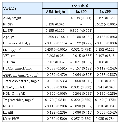
Fourteen patients (13.7%) were diagnosed with impaired microcirculatory function of the lower limb based on SPP. The prevalence of sarcopenia in all subjects was 11.8%, but the percentage of patients with an SPP ≤50 mm Hg who had sarcopenia was more than triple that of patients with an SPP >50 mm Hg (28.6% vs. 9.1%, P=0.036). A significant positive correlation was found between SPP and appendicular muscle mass adjusted for height (P=0.041 for right-sided SPP). Multiple logistic regression analysis showed that patients with sarcopenia had an odds ratio of 4.1 (95% confidence interval, 1.01 to 24.9) for having an SPP ≤50 mm Hg even after adjustment for confounding factors.
Conclusion
These results suggest that sarcopenia may be significantly associated with impaired microcirculation in patients with T2DM. Nonetheless, the small number of patients and wide CI require cautious interpretation of the results. -
Citations
Citations to this article as recorded by
- Preclinical study of diabetic foot ulcers: From pathogenesis to vivo/vitro models and clinical therapeutic transformation
Yuqing Du, Jie Wang, Weijing Fan, Renyan Huang, Hongfei Wang, Guobin Liu
International Wound Journal.2023; 20(10): 4394. CrossRef - Bioelectrical Impedance Analysis for the Assessment of Body Composition in Sarcopenia and Type 2 Diabetes
Stefano Sbrignadello, Christian Göbl, Andrea Tura
Nutrients.2022; 14(9): 1864. CrossRef - Discrimination between possible sarcopenia and metabolic syndrome using the arterial pulse spectrum and machine-learning analysis
Li-Wei Wu, Te OuYoung, Yu-Chih Chiu, Ho-Feng Hsieh, Hsin Hsiu
Scientific Reports.2022;[Epub] CrossRef - The prevalence and risk factors of sarcopenia in patients with type 2 diabetes mellitus: a systematic review and meta-analysis
Yaqin Ai, Ruoxin Xu, Lingping Liu
Diabetology & Metabolic Syndrome.2021;[Epub] CrossRef
- Preclinical study of diabetic foot ulcers: From pathogenesis to vivo/vitro models and clinical therapeutic transformation

- Ca Effects on Synthesis and Secretion of Insulin-like Growth Factor(IGF-I) and IGF-Binding Proteins by the Perfased Rat Liver.
- Dae Yeol Lee, Chang Won Kang, Jung Soo Kim
- J Korean Endocr Soc. 1996;11(2):189-198. Published online November 7, 2019
- 1,248 View
- 26 Download
-
Abstract
PDF
- Background
The insulin-like growth factors, IGF-I and -II, are important metabolic factors involved in cell growth and metabolism. The IGFs are produced in most organs but the liver is believed to be the principal source of circulating IGF-I. It has been demonstrated that calcium in the extracellular fluid has effects on the secretion of various hormones such as parathyroid hormone, insulin and atrial natriuretic peptide in variety of tissues. Methods: In arder to investigate that liver produce IGF-1 and IGFBP-3 and the role of calcium in the regulation of IGF-I and IGFBP-3 secretion and synthesis, the rat liver perfusian model was employed. The liver was perfused with Krebs-Henseleit bicarbonate(KHB) buffer containing 0 or 2.5 mM CaC12 for 2 hours, and 4-ml fractions of perfusates were collected and determined the concentration of IGF-I and IGFBP-3 by using RIA after Sep-pak extraction and Western ligand blot. respectively. To increase or decrease the concentration of intracellular calcium, we also used EGTA, calciurn ionophore A23187 increased IGF-I secretion and synthesis in liver(18.13+0.97 vs 15.78+1.01, p<0.01). However, concentration of glucose was not significantly affected by both calcium(2.07+1.44 vs 2.24+1.74) and BGTA(2.01+1A7 vs 3.11+1.01). Conclusion: Our results demonstrate that liver is a major place far IGF-I and IOFBP-3 production and calcium specifically affects the secretion of IGF-I in the liver perfusion, suggesting that the calcium environment of hepatic cells may influence the secretion of the hepatic IGF-I.
 First
First Prev
Prev


