Articles
- Page Path
- HOME > Endocrinol Metab > Volume 39(1); 2024 > Article
-
Review ArticleThyroid Novel and Advanced Ultrasound Techniques for Thyroid Thermal Ablation
 Keypoint
Thyroid radiofrequency ablation and microwave ablation are widely adopted minimally invasive treatments for diverse thyroid conditions worldwide. This review discusses the basic techniques and mechanisms of thyroid thermal ablation. Moreover, this review introduces essential maneuvers, including the zigzag moving technique, hydrodissection, the alienate maneuver, and vascular ablation. -
Wai-Kin Chan1
 , Jui-Hung Sun1, Miaw-Jene Liou1,2, Chia-Jung Hsu1, Yu-Ling Lu1, Wei-Yu Chou1, Yan-Rong Li1,2, Feng-Hsuan Liu1,2
 -
Endocrinology and Metabolism 2024;39(1):40-46.
DOI: https://doi.org/10.3803/EnM.2024.1917
Published online: February 13, 2024
1Division of Endocrinology and Metabolism, Department of Internal Medicine, Chang Gung Memorial Hospital, Taoyuan, Taiwan
2Chang Gung University, College of Medicine, Taoyuan, Taiwan
- Corresponding author: Feng-Hsuan Liu. Division of Endocrinology and Metabolism, Department of Internal Medicine, Chang Gung Memorial Hospital, Chang Gung University, College of Medicine, No. 5, Fuxing St., Guishan Dist., Taoyuan, Taiwan Tel: +886-975368146, Fax: +886-3-3285060, E-mail: a122liu@cgmh.org.tw
• Received: December 29, 2023 • Revised: January 9, 2024 • Accepted: January 15, 2024
Copyright © 2024 Korean Endocrine Society
This is an Open Access article distributed under the terms of the Creative Commons Attribution Non-Commercial License (http://creativecommons.org/licenses/by-nc/4.0/) which permits unrestricted non-commercial use, distribution, and reproduction in any medium, provided the original work is properly cited.
- 1,717 Views
- 114 Download
ABSTRACT
- Thyroid radiofrequency ablation and microwave ablation are widely adopted minimally invasive treatments for diverse thyroid conditions worldwide. Fundamental skills such as the trans-isthmic approach and the moving shot technique are crucial for performing thyroid ablation, and advanced techniques, including hydrodissection and vascular ablation, improve safety and efficacy and reduce complications. Given the learning curve associated with ultrasound-guided therapeutic procedures, operators need training and experience. While training models exist, limited attention has been given to ultrasound maneuvers in ablation needle manipulation. This article introduces two essential maneuvers, the zigzag moving technique and the alienate maneuver, while also reviewing the latest ultrasound techniques in thyroid ablation, contributing valuable insights into this evolving field.
- Thyroid radiofrequency ablation (RFA) [1] and microwave ablation (MWA) [2] are novel minimally invasive treatments that have gained widespread adoption worldwide. They have been shown to be effective and safe for managing benign thyroid nodules, cystic nodules, parathyroid adenoma, papillary thyroid microcarcinoma, metastatic lymph nodes, and inoperable recurrent thyroid cancers. After ablation, these thyroid lesions typically manifest with firm and heterogeneous hypoechogenicity on ultrasonography (Fig. 1). Free-handed ultrasound (US) techniques, including the trans-isthmic approach [3,4] and the moving shot technique [5,6], have become fundamental skills for performing thyroid ablation. Moreover, advanced US techniques, such as hydrodissection (HD) and vascular ablation, have been introduced to improve safety and efficacy, thereby minimizing complications [7]. It is important to recognize that there is a learning curve associated with US-guided therapeutic procedures [8-10], and operators undertaking thyroid ablation require appropriate training and experience. While a successful training model [11] has been introduced to expedite the learning curve, limited attention has been given to training on US maneuvers in manipulating the ablation needle. This article aims to introduce two fundamental maneuvers, namely the zigzag moving technique and alienate maneuver. Additionally, we review the latest US techniques employed in thyroid ablation procedures.
INTRODUCTION
- There have been numerous advances in US techniques for treating thyroid nodules (Table 1). The trans-isthmic approach and moving shot technique have become basic and core techniques in performing thyroid ablation [12]. Caution is needed to avoid damage to the critical structures surrounding the thyroid gland, including nerves, large vessels, and the esophagus [13,14]. The trans-isthmic approach is specifically employed to prevent damage to the “danger triangle,” which includes the recurrent laryngeal nerve (RLN), which is located between the tracheoesophageal groove and the thyroid gland. This approach involves inserting the electrode needle through the midline to the lateral aspect of the neck. Local anesthesia is administered between the thyroid capsule and adjacent muscles to minimize pain during treatment. For nodules located close to critical structures, HD techniques have emerged as advanced yet core approaches for separating the nodule from these structures. Experienced operators may consider various approaches and needle retention methods during HD [15], such as the antero-lateral approach, danger triangle approach, pre-tracheal approach, and posterior approach. In the treatment planning of nodule ablation, the target nodule is typically divided into several conceptual units for ablation based on the nodule’s size [1]. The free-handed moving shot technique is recommended over the fixed electrode technique [1,16], as it allows higher ablation efficacy, facilitating the complete ablation of all units and enabling the delivery of increased ablation energy.
- In RFA, the generation of frictional heat results from agitated ions with a local temperature increase induced by high-frequency alternating electric current flowing around the electrode. This process causes protein denaturation and tissue necrosis. Greater ablation volume and more extensive tissue damage can occur through thermal radiation and conduction whenever the active tip is fixed within one area [1]. Modern RFA devices are designed to minimize tissue carbonization by employing internal water cooling in the center of the electrodes, maintaining a steady temperature of around 60°C during ablation. In contrast, MWA utilizes microwaves to heat water molecules, ensuring a consistent power output unaffected by electrical impedance, thereby reducing treatment time. Furthermore, the technical US requirements for both devices are nearly identical.
- The zigzag moving technique
- We introduce a crucial maneuver called the “zigzag moving technique” for manipulating the RFA electrode or MWA antenna while transitioning between ablation units within the target lesion. Typically, after the insertion of the electrode through the trans-isthmic approach under US guidance, ablation begins from the deepest and most remote portion of the nodule. The needle tip is then gradually retracted toward the margin of the nodule. This process is repeated with a less tilted angle towards the superficial portion until the entire unit is completely treated. Upon the complete ablation of the current unit, the electrode tip is drawn back to the edge of the nodule, ideally without exiting the nodule.
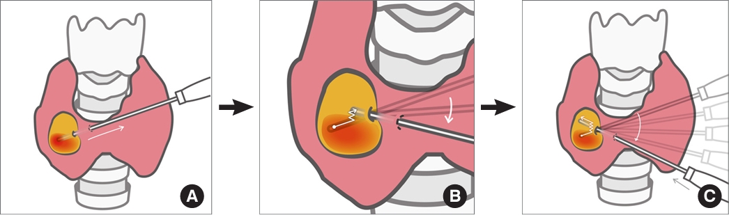
- To access the next intended ablation unit, a “zigzag” or “sawtooth”-like movement of the tip toward the next unit is recommended (Fig. 2). Simultaneously, the position of the US probe is adjusted with a wedge-shaped movement to maintain alignment and visualize the electrode tip during each move. This technique aims to minimize puncture holes within the ablated nodule and is strongly recommended for all operators to incorporate into their thyroid ablation procedures. For beginners, mastering this technique prevents disorientation regarding the position of the electrode/needle tip and may lead to a faster learning curve, allowing them to achieve larger ablation volumes in less time.
BASIC TECHNIQUES AND MECHANISMS OF THYROID THERMAL ABLATION
- Hydrodissection
- The HD technique is extensively employed in both RFA and MWA to ablate liver and thyroid tumors. It has proven to be an effective method for separating the target lesion from critical structures by creating a liquid buffer [12,14]. Various methods can be utilized for HD. One of the fundamental US techniques is the perithyroidal lidocaine injection, also known as the anterolateral approach HD (Fig. 3) [17]. This technique aims to minimize pain during ablation and to protect adjacent structures, especially those located laterally to the thyroid, such as the middle cervical sympathetic ganglion and the vagus nerve [15,18,19]. The injected lidocaine serves as a temporary barrier between the thyroid capsule, strap muscles, and carotid sheath, but it eventually disperses into the mediastinum [20]. Care should be taken to avoid complications such as voice change if lidocaine leaks into the peri-tracheal area. A 5% dextrose solution is the preferred medium for HD over normal saline because normal saline, as an ionic liquid, can conduct electricity and potentially cause damage to nearby structures through friction heat [1,13]. However, this concern is not applicable to MWA, making normal saline the preferred choice for this method. The “danger triangle” approach is a commonly used HD technique (Fig. 4), where the solution is injected from the posterior surface of the isthmus (pre-tracheal) and directed toward the space around the “danger triangle.” Skilled practitioners may choose to keep the injection needle in place (Fig. 5) [17,20] to continuously administer the solution and maintain the dissection gap. The posterior approach, involving HD between the thyroid and retropharyngeal space, can be applied using either trans-isthmic or lateral cervical approaches (Fig. 6) [15]. The posterior approach is strongly recommended for ablating deep nodules in the right lobe, considering the slightly lateral path of the RLN compared to the left side [10]. Using a large volume of HD fluid, typically greater than 40 mL, has yielded satisfactory outcomes [20]. In our experience, transient hoarseness or voice changes can sometimes occur due to compression and the cooling effect on the superior laryngeal nerve or RLN, but these symptoms usually resolve before the ablation procedure is completed.
- The alienate maneuver
- In addition to HD, we introduce another concept, termed the “alienate maneuver.” In scenarios involving the trans-isthmic approach, once the needle has been advanced to the deepest and most remote part of the nodule, with the isthmus serving as a pivot, firm and steady upward pressure can be exerted toward the antero-lateral aspect of the neck. The purpose of this maneuver is to elevate the nodule, moving it away from the danger triangle and the posterior compartment, allowing for complete ablation of the deepest portion using the moving shot technique. For this maneuver, it is preferable to use electrodes or antennas with a larger diameter (16 to 18 gauge), as thinner needles may bend when force is applied. Theoretically, the combined use of HD and the alienate maneuver could further minimize the risk of thermal injury (Fig. 7).
TECHNIQUES FOR AVOIDING THERMAL INJURIES
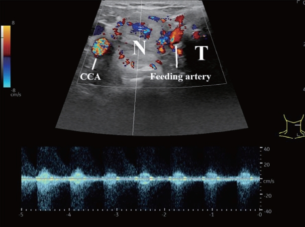
- Artery-first ablation and marginal venous ablation [7,21] are two recommended techniques aimed at reducing the heat-sink effect [22], decreasing the risk of hemorrhage, and minimizing potential marginal growth. Although arterial ablation is not required for every patient and locating the feeding vessels can be challenging in some instances, it is advantageous when possible. In hyper-vascular thyroid nodules, a prominent feeding artery is often detectable using spectral Doppler imaging (Fig. 8), typically originating from either the superior or inferior thyroid artery. If a feeding artery is ablated, intra-nodular linear hyperechogenicity caused by microbubbles within the supplying vessels or a wedge-shaped hypodense region representing infarction of the corresponding part may be observed. Patients may experience pain during artery ablation due to innervation within the arterial walls.
- The marginal venous ablation technique is widely applied, given that most thyroid nodules have peri-nodular veins. Similar to the ablation of arteries, this technique results in vessels filled with microbubbles, displaying hyperechogenicity at the superficial part of the nodule (Fig. 9). When both techniques are employed concurrently, arterial ablation should precede venous ablation. Previous studies suggest that the marginal venous ablation technique may be beneficial for preventing marginal regrowth [7,23,24].
VASCULAR ABLATION
- The zigzag moving technique is a recommended manipulation for controlling the RFA electrode or MWA antenna during the transition between conceptual ablation treatment units within a nodule. It is important for operators to understand and practice these fundamental and advanced US techniques, including the alienate maneuver, HD, and vascular ablation, to ensure a safe and effective treatment.
CONCLUSIONS
-
CONFLICTS OF INTEREST
No potential conflict of interest relevant to this article was reported.
Article information
-
Acknowledgements
- This study was supported by Chang Gung Medical Research Project (CMRP) for research funding (CMRPG3N0811). The authors wish to thank Professor Hong-Cheng Wang for his invaluable suggestions and opinions.
Fig. 1.(A) Pre-radiofrequency ablation (RFA) ultrasound of thyroid nodule. (B) Post-RFA ultrasound of the same thyroid nodule (arrows). CCA, common carotid artery; T, trachea.
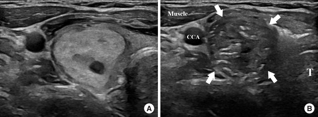
Fig. 2.Illustrations of the procedure for the zigzag moving technique from a front view. (A) After ablating the lower unit, the needle tip is withdrawn to the periphery of the nodule. (B) The needle is then maneuvered within the nodule in a zigzag or sawtooth-like pattern towards the upper ablation unit (the path of the needle tip is indicated by the white line). (C) This movement is continued until the desired ultrasound plane is accurately reached. Subsequently, the electrode is advanced to the deepest part of the target area to begin ablation of the upper unit (the white line depicts the complete trajectory of the needle).

Fig. 3.Perithyroidal lidocaine injection and antero-lateral hydrodissection with a retained needle for continuous hydrodissection. CCA, common carotid artery; T, trachea.
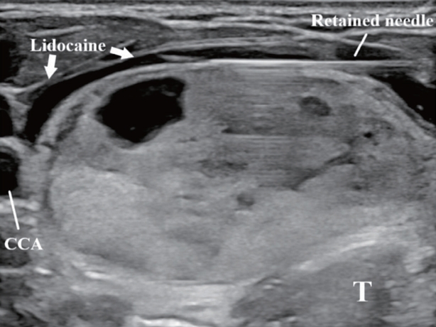
Fig. 4.(A) Ultrasonography of a high-risk thyroid nodule (N) adjacent to the trachea (T) and danger triangle. A perithyroidal lidocaine injection was performed (arrows). (B) Hydrodissection by the danger triangle approach with a continuous injection of 5% dextrose solution (arrows). A sufficient safety margins between the N and peri-tracheal region was secured. CCA, common carotid artery.
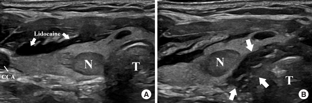
Fig. 5.Image illustrating the actual performance of continuous hydrodissection and radiofrequency ablation. A 21G needle was connected to a dextrose solution syringe by the danger triangle approach through the isthmus (arrow). US, ultrasound; D5W, 5% dextrose water.
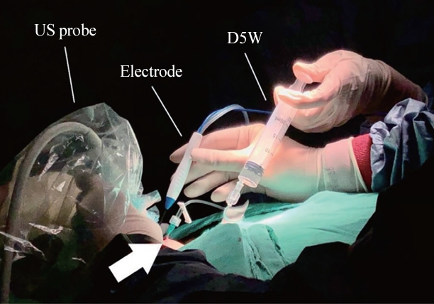
Fig. 6.Hydrodissection by the posterior approach with a continuous injection. With a large enough amount of hydrodissection solution, both the lateral compartment and posterior compartment were covered. CCA, common carotid artery; N, nodule; T, trachea.
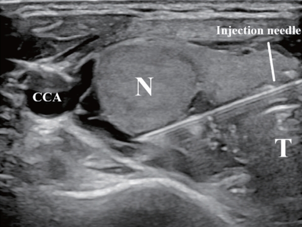
Fig. 7.The alienate maneuver. After advancing the electrode to the deepest portion of the nodule (N), upward pressure (arrow) can be applied towards the antero-lateral part of the neck. With the help of antero-lateral and danger triangle hydrodissection (HD), the N was isolated from critical structures. CCA, common carotid artery; T, trachea.
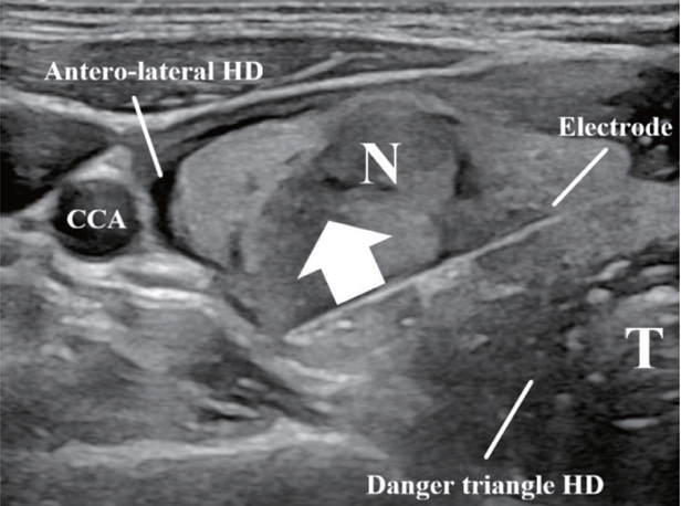
Fig. 8.Feeding vessel entering a hyper-vascular nodule (N). The feeding artery can be distinguished and located by color and spectral Doppler showing an arterial waveform. CCA, common carotid artery; T, trachea.

Fig. 9.Ultrasound of a hyper-vascular nodule (N) after marginal vein ablation. Hyperechoic air bubbles (arrows) fill the veins and can be observed at the superficial part of the ablated N. CCA, common carotid artery; T, trachea.

Table 1.Summary of Basic and Advanced Ultrasound Techniques
- 1. Shin JH, Baek JH, Ha EJ, Lee JH. Radiofrequency ablation of thyroid nodules: basic principles and clinical application. Int J Endocrinol 2012;2012:919650.ArticlePubMedPMCPDF
- 2. Morelli F, Sacrini A, Pompili G, Borelli A, Panella S, Masu A, et al. Microwave ablation for thyroid nodules: a new string to the bow for percutaneous treatments? Gland Surg 2016;5:553–8.ArticlePubMedPMC
- 3. Ahn HS, Kim SJ, Park SH, Seo M. Radiofrequency ablation of benign thyroid nodules: evaluation of the treatment efficacy using ultrasonography. Ultrasonography 2016;35:244–52.ArticlePubMedPMCPDF
- 4. Baek JH, Moon WJ, Kim YS, Lee JH, Lee D. Radiofrequency ablation for the treatment of autonomously functioning thyroid nodules. World J Surg 2009;33:1971–7.ArticlePubMedPDF
- 5. Ha EJ, Baek JH, Lee JH. Moving-shot versus fixed electrode techniques for radiofrequency ablation: comparison in an exvivo bovine liver tissue model. Korean J Radiol 2014;15:836–43.ArticlePubMedPMC
- 6. Jeong WK, Baek JH, Rhim H, Kim YS, Kwak MS, Jeong HJ, et al. Radiofrequency ablation of benign thyroid nodules: safety and imaging follow-up in 236 patients. Eur Radiol 2008;18:1244–50.ArticlePubMedPDF
- 7. Park HS, Baek JH, Park AW, Chung SR, Choi YJ, Lee JH. Thyroid radiofrequency ablation: updates on innovative devices and techniques. Korean J Radiol 2017;18:615–23.ArticlePubMedPMCPDF
- 8. Kuo CY, Liu CL, Tsai CH, Cheng SP. Learning curve analysis of radiofrequency ablation for benign thyroid nodules. Int J Hyperthermia 2021;38:1536–40.ArticlePubMed
- 9. Russ G, Ben Hamou A, Poiree S, Ghander C, Menegaux F, Leenhardt L, et al. Learning curve for radiofrequency ablation of benign thyroid nodules. Int J Hyperthermia 2021;38:55–64.ArticlePubMed
- 10. Sinclair CF, Baek JH, Hands KE, Hodak SP, Huber TC, Hussain I, et al. General principles for the safe performance, training, and adoption of ablation techniques for benign thyroid nodules: an American Thyroid Association Statement. Thyroid 2023;33:1150–70.ArticlePubMedPMC
- 11. Li YR, Chou WY, Chan WK, Cheng KL, Sun JH, Liu FH, et al. Successful applications of food-assisted and -simulated training model of thyroid radiofrequency ablation. Front Endocrinol (Lausanne) 2022;13:809835.ArticlePubMedPMC
- 12. Ha EJ, Baek JH, Che Y, Chou YH, Fukunari N, Kim JH, et al. Radiofrequency ablation of benign thyroid nodules: recommendations from the Asian Conference on Tumor Ablation Task Force. Ultrasonography 2021;40:75–82.ArticlePubMedPMCPDF
- 13. Kim JH, Baek JH, Lim HK, Ahn HS, Baek SM, Choi YJ, et al. 2017 Thyroid radiofrequency ablation guideline: Korean Society of Thyroid Radiology. Korean J Radiol 2018;19:632–55.ArticlePubMedPMCPDF
- 14. Xiaoyin T, Ping L, Dan C, Min D, Jiachang C, Tao W, et al. Risk assessment and hydrodissection technique for radiofrequency ablation of thyroid benign nodules. J Cancer 2018;9:3058–66.ArticlePubMedPMC
- 15. Chung SR, Baek JH, Choi YJ, Lee JH. Thermal ablation for the management of papillary thyroid microcarcinoma in the era of active surveillance and hemithyroidectomy. Curr Oncol Rep 2022;24:1045–52.ArticlePubMedPDF
- 16. Baek JH, Kim YS, Lee D, Huh JY, Lee JH. Benign predominantly solid thyroid nodules: prospective study of efficacy of sonographically guided radiofrequency ablation versus control condition. AJR Am J Roentgenol 2010;194:1137–42.ArticlePubMed
- 17. Jeong SY, Baek JH, Chung SR, Choi YJ, Chung KW, Kim TY, et al. Radiofrequency ablation of benign thyroid nodules: the value of anterolateral hydrodissection. Ultrasonography 2023;42:432–9.ArticlePubMedPMCPDF
- 18. Chung SR, Suh CH, Baek JH, Park HS, Choi YJ, Lee JH. Safety of radiofrequency ablation of benign thyroid nodules and recurrent thyroid cancers: a systematic review and meta-analysis. Int J Hyperthermia 2017;33:920–30.ArticlePubMed
- 19. Cho SJ, Baek JH, Chung SR, Choi YJ, Lee JH. Long-term results of thermal ablation of benign thyroid nodules: a systematic review and meta-analysis. Endocrinol Metab (Seoul) 2020;35:339–50.ArticlePubMedPMCPDF
- 20. Ma Y, Wu T, Yao Z, Zheng B, Tan L, Tong G, et al. Continuous, large-volume hydrodissection to protect delicate structures around the thyroid throughout the radiofrequency ablation procedure. Eur Thyroid J 2021;10:495–503.ArticlePubMedPMCPDF
- 21. Lee M, Baek JH, Suh CH, Chung SR, Choi YJ, Lee JH, et al. Clinical practice guidelines for radiofrequency ablation of benign thyroid nodules: a systematic review. Ultrasonography 2021;40:256–64.ArticlePubMedPMCPDF
- 22. Pillai K, Akhter J, Chua TC, Shehata M, Alzahrani N, AlAlem I, et al. Heat sink effect on tumor ablation characteristics as observed in monopolar radiofrequency, bipolar radiofrequency, and microwave, using ex vivo calf liver model. Medicine (Baltimore) 2015;94:e580.ArticlePubMedPMC
- 23. Wang B, Han ZY, Yu J, Cheng Z, Liu F, Yu XL, et al. Factors related to recurrence of the benign non-functioning thyroid nodules after percutaneous microwave ablation. Int J Hyperthermia 2017;33:459–64.ArticlePubMed
- 24. Zhao CK, Xu HX, Lu F, Sun LP, He YP, Guo LH, et al. Factors associated with initial incomplete ablation for benign thyroid nodules after radiofrequency ablation: first results of CEUS evaluation. Clin Hemorheol Microcirc 2017;65:393–405.ArticlePubMed
References
Figure & Data
References
Citations
Citations to this article as recorded by 
 PubReader
 ePub Link
 Cite
Cite