
Articles
- Page Path
- HOME > Endocrinol Metab > Volume 29(3); 2014 > Article
-
Original ArticleEndocrine Research Polarized and Stage-Dependent Distribution of Immunoreactivity for Novel PDZ-Binding Protein Preso1 in Adult Neurogenic Regions
- Eun Soo Lee1*, Woon Ryoung Kim1*, Younghwa Kim2, Hyun Woo Lee1, Hyun Kim1, Woong Sun1
-
Endocrinology and Metabolism 2014;29(3):349-355.
DOI: https://doi.org/10.3803/EnM.2014.29.3.349
Published online: September 25, 2014
1Department of Anatomy, BK21 Program, Korea University College of Medicine, Seoul, Korea.
2Department of Emergency Medical Technology, Kyungil University College of Nursing and Public Health, Gyeongsan, Korea.
- Corresponding author: Woong Sun. Department of Anatomy, Korea University College of Medicine, 73 Inchon-ro, Seongbuk-gu, Seoul 136-705, Korea. Tel: +82-2-920-6404, Fax: +82-2-929-5696, woongsun@korea.ac.kr
- *These authors contributed equally to this work.
• Received: September 28, 2013 • Revised: November 3, 2013 • Accepted: January 13, 2014
Copyright © 2014 Korean Endocrine Society
This is an Open Access article distributed under the terms of the Creative Commons Attribution Non-Commercial License (http://creativecommons.org/licenses/by-nc/3.0/) which permits unrestricted non-commercial use, distribution, and reproduction in any medium, provided the original work is properly cited.
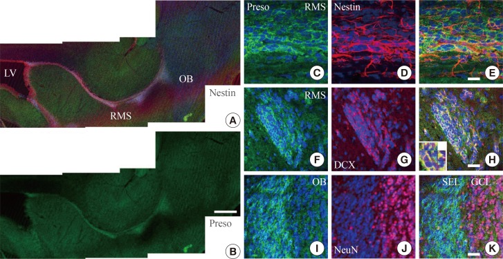
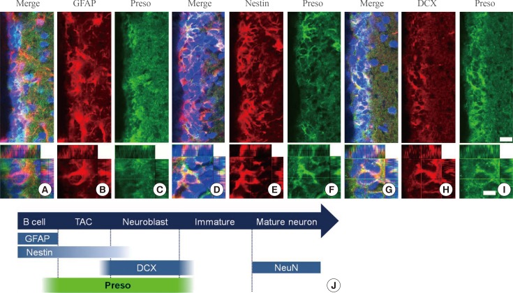
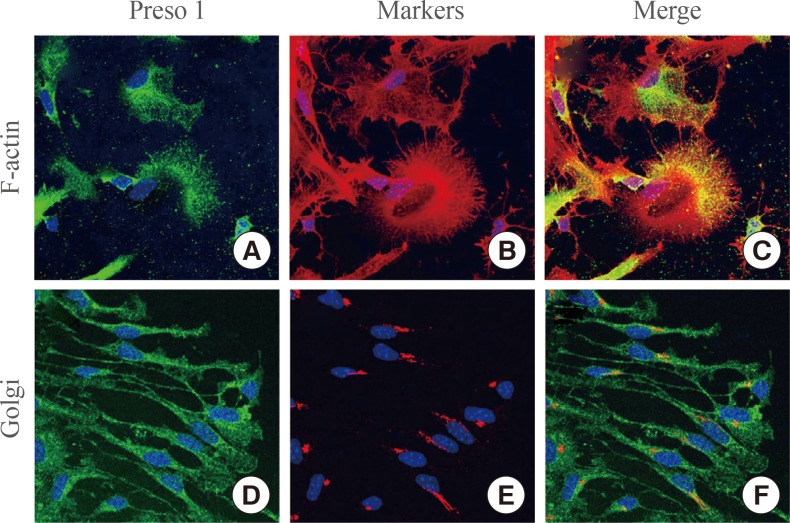
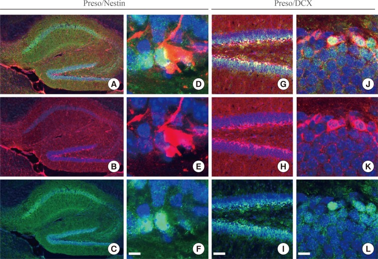
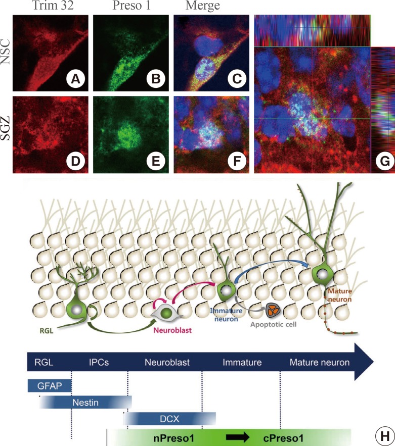
Figure & Data
References
Citations
Citations to this article as recorded by 
- FERM domain–containing proteins are active components of the cell nucleus
Péter Borkúti, Ildikó Kristó, Anikó Szabó, Zoltán Kovács, Péter Vilmos
Life Science Alliance.2024; 7(4): e202302489. CrossRef - Articles in 'Endocrinology and Metabolism' in 2014
Won-Young Lee
Endocrinology and Metabolism.2015; 30(1): 47. CrossRef
 PubReader
 Cite
Cite


